[English] 日本語
Yorodumi
- PDB-2hn1: Crystal structure of a CorA soluble domain from A. fulgidus in co... -

+
Open data


ID or keywords:

Loading...

-
Basic information

Entry
Database: PDB / ID: 2hn1
TitleCrystal structure of a CorA soluble domain from A. fulgidus in complex with Co2+
ComponentsMagnesium and cobalt transporter
KeywordsMETAL TRANSPORT / integral membrane protein fragment / metal transporter protein / divalent cations
Function / homology
Function and homology information


cobalt ion transmembrane transporter activity / magnesium ion transmembrane transporter activity / membrane => GO:0016020 / plasma membrane
Similarity search - Function
Magnesium transport protein CorA, transmembrane region / CorA soluble domain-like / Magnesium/cobalt transport protein CorA / Beta Polymerase; domain 2 / Methane Monooxygenase Hydroxylase; Chain G, domain 1 / Up-down Bundle / 2-Layer Sandwich / Mainly Alpha / Alpha Beta
Similarity search - Domain/homology
: / Magnesium transport protein CorA
Similarity search - Component
Biological speciesArchaeoglobus fulgidus (archaea)
MethodX-RAY DIFFRACTION / SYNCHROTRON / MAD / Resolution: 2.9 Å
AuthorsPayandeh, J. / Pai, E.F.
CitationJournal: Embo J. / Year: 2006
Title: A structural basis for Mg(2+) homeostasis and the CorA translocation cycle.
Authors: Payandeh, J. / Pai, E.F.
History
DepositionJul 11, 2006Deposition site: RCSB / Processing site: RCSB
Revision 1.0Aug 29, 2006Provider: repository / Type: Initial release
Revision 1.1May 1, 2008Group: Version format compliance
Revision 1.2Jul 13, 2011Group: Derived calculations / Version format compliance
Revision 1.3Oct 18, 2017Group: Refinement description / Category: software
Item: _software.classification / _software.contact_author ..._software.classification / _software.contact_author / _software.contact_author_email / _software.date / _software.language / _software.location / _software.name / _software.type / _software.version
Revision 1.4Feb 14, 2024Group: Data collection / Database references / Derived calculations
Category: chem_comp_atom / chem_comp_bond ...chem_comp_atom / chem_comp_bond / database_2 / pdbx_struct_conn_angle / struct_conn / struct_ref_seq_dif / struct_site
Item: _database_2.pdbx_DOI / _database_2.pdbx_database_accession ..._database_2.pdbx_DOI / _database_2.pdbx_database_accession / _pdbx_struct_conn_angle.ptnr1_auth_comp_id / _pdbx_struct_conn_angle.ptnr1_auth_seq_id / _pdbx_struct_conn_angle.ptnr1_label_atom_id / _pdbx_struct_conn_angle.ptnr1_label_comp_id / _pdbx_struct_conn_angle.ptnr1_label_seq_id / _pdbx_struct_conn_angle.ptnr1_symmetry / _pdbx_struct_conn_angle.ptnr3_auth_comp_id / _pdbx_struct_conn_angle.ptnr3_auth_seq_id / _pdbx_struct_conn_angle.ptnr3_label_atom_id / _pdbx_struct_conn_angle.ptnr3_label_comp_id / _pdbx_struct_conn_angle.ptnr3_label_seq_id / _pdbx_struct_conn_angle.ptnr3_symmetry / _pdbx_struct_conn_angle.value / _struct_conn.pdbx_dist_value / _struct_conn.ptnr1_auth_comp_id / _struct_conn.ptnr1_auth_seq_id / _struct_conn.ptnr1_label_asym_id / _struct_conn.ptnr1_label_atom_id / _struct_conn.ptnr1_label_comp_id / _struct_conn.ptnr1_label_seq_id / _struct_conn.ptnr1_symmetry / _struct_conn.ptnr2_auth_comp_id / _struct_conn.ptnr2_auth_seq_id / _struct_conn.ptnr2_label_asym_id / _struct_conn.ptnr2_label_atom_id / _struct_conn.ptnr2_label_comp_id / _struct_conn.ptnr2_label_seq_id / _struct_conn.ptnr2_symmetry / _struct_ref_seq_dif.details / _struct_site.pdbx_auth_asym_id / _struct_site.pdbx_auth_comp_id / _struct_site.pdbx_auth_seq_id
Remark 300 
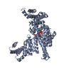
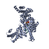
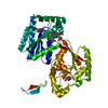
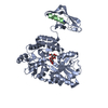
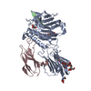
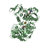
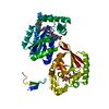
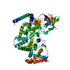
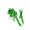
BIOMOLECULE: 1 THE BIOLOGICALLY RELEVANT MOLECULE OF FULL-LENGTH CORA IS A HOMOPENTAMER. HERE, WE ... BIOMOLECULE: 1 THE BIOLOGICALLY RELEVANT MOLECULE OF FULL-LENGTH CORA IS A HOMOPENTAMER. HERE, WE FIND A SINGLE MOLECULE OF THE SOLUBLE DOMAIN WITHIN THE ASSYMETRIC UNIT, AND A DOMAIN-SWAPPED DIMER IS FORMED BY SPACE GROUP SYMMETRY. WE HAVE PROPOSED THAT THE CONFORMATIONAL CHANGE OBSERVED HERE, RELATIVE TO THE FULL-LENGTH PROTEIN, MAY INDICATE A MECHANISM FOR GATING IN THE FULL-LENGTH TRANSPORTER PROTEIN.

-
Structure visualization

Structure viewerMolecule:
MolmilJmol/JSmol

Downloads & links

-
Assembly

Deposited unit
A: Magnesium and cobalt transporter
hetero molecules


Theoretical massNumber of molelcules
Total (without water)30,5372
Polymers30,4781
Non-polymers591
Water00
1
A: Magnesium and cobalt transporter
hetero molecules

A: Magnesium and cobalt transporter
hetero molecules


Theoretical massNumber of molelcules
Total (without water)61,0744
Polymers60,9562
Non-polymers1182
Water0
TypeNameSymmetry operationNumber
identity operation1_555x,y,z1
crystal symmetry operation8_555x-y,-y,-z1
Buried area7590 Å2
ΔGint-65 kcal/mol
Surface area23620 Å2
MethodPISA, PQS
Unit cell
Length a, b, c (Å)101.216, 101.216, 142.724
Angle α, β, γ (deg.)90.000, 90.000, 120.000
Int Tables number178
Space group name H-MP6122
DetailsThe biologically relavent molecule of CorA is a pentamer. A single chain is observed in the assymetric unit, and a domain-swapped dimer is formed by space group symmetry. When compared to other known structures of CorA, the conformational changes observed in this soluble domain structure may indicate a mechanism for activation (i.e. CorA gating). Alternatively, the conformation observed could be a cloning and/or crystallization artifact. The Co2+ coordinated to Asp253 and His257 was confirmed experimentally by solving this structure independently from a MAD experiment (performed on a native crystal) at the cobalt absorption edge.

-
Components

#1: Protein Magnesium and cobalt transporter / CorA


Mass: 30478.227 Da / Num. of mol.: 1 / Fragment: N-terminal soluble domain
Source method: isolated from a genetically manipulated source
Source: (gene. exp.) Archaeoglobus fulgidus (archaea) / Strain: ATCC 49558D / Gene: CorA / Plasmid: pET15-b / Production host: Escherichia coli (E. coli) / Strain (production host): BL21-CodonPlus(DE3)-RIL / References: UniProt: O29472
#2: Chemical ChemComp-CO / COBALT (II) ION


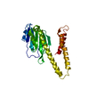
Mass: 58.933 Da / Num. of mol.: 1 / Source method: obtained synthetically / Formula: Co

-
Experimental details

-
Experiment

ExperimentMethod: X-RAY DIFFRACTION / Number of used crystals: 1

-
Sample preparation

CrystalDensity Matthews: 3.46 Å3/Da / Density % sol: 64.47 %
Crystal growTemperature: 295 K / Method: vapor diffusion, hanging drop / pH: 7.4
Details: 1.3 M ammonium sulfate, 10 mM CoCl2, 0.1 M HEPES pH 7.4, VAPOR DIFFUSION, HANGING DROP, temperature 295K

-
Data collection

Diffraction
IDMean temperature (K)Crystal-ID
11001
21
31
Diffraction sourceSource: SYNCHROTRON / Site: NSLS / Beamline: X8C / Wavelength: 1.1 Å
DetectorType: ADSC QUANTUM 4 / Detector: CCD / Date: Sep 24, 2005
RadiationProtocol: SINGLE WAVELENGTH / Monochromatic (M) / Laue (L): M / Scattering type: x-ray
Radiation wavelengthWavelength: 1.1 Å / Relative weight: 1
Reflection

D res high: 3.2 Å / D res low: 100 Å

IDAv σ(I) over netINumberRmerge(I) obsΧ2Num. obs% possible obs
127.71748330.0871.36761899.9
230.51758940.0751.19765899.8
329.31756990.0731.28765299.9
Diffraction reflection shell
Highest resolution (Å)Lowest resolution (Å)% possible obs (%)IDRmerge(I) obsChi squared
6.8910099.110.0692.109
5.476.8910010.0742.114
4.785.4710010.0711.826
4.344.7810010.0681.608
4.034.3410010.081.118
3.794.0310010.1091.084
3.63.7910010.1481.069
3.453.610010.2020.942
3.313.4510010.2890.87
3.23.3110010.4230.853
6.8910098.520.0581.769
5.476.8910020.0631.818
4.785.4710020.061.507
4.344.7810020.0581.318
4.034.3410020.0680.965
3.794.0310020.0950.984
3.63.7910020.130.97
3.453.610020.180.883
3.313.4510020.2650.834
3.23.3110020.3930.821
6.8910098.930.0551.904
5.476.8910030.0591.886
4.785.4710030.0571.612
4.344.7810030.0561.486
4.034.3410030.0681.153
3.794.0310030.0951.026
3.63.7910030.1371.024
3.453.610030.1890.943
3.313.4510030.2750.877
3.23.3110030.4110.859
ReflectionResolution: 2.9→100 Å / Num. all: 10178 / Num. obs: 9953 / % possible obs: 100 % / Observed criterion σ(I): 2 / Rmerge(I) obs: 0.078 / Χ2: 1.392 / Net I/σ(I): 24.5
Reflection shellResolution: 2.9→3 Å / Rmerge(I) obs: 0.479 / Num. unique all: 977 / Χ2: 1.211 / % possible all: 100

-
Phasing

PhasingMethod: MAD
Phasing set
ID
1
2
3
Phasing MAD set
Clust-IDExpt-IDSet-IDWavelength (Å)F double prime refinedF prime refined
13 wavelength10.97944.98-8.15
13 wavelength20.98012.79-9.55
13 wavelength30.963.7-3.2
Phasing dmFOM : 0.72 / FOM acentric: 0.72 / FOM centric: 0.73 / Reflection: 7413 / Reflection acentric: 5828 / Reflection centric: 1585
Phasing dm shell
Resolution (Å)FOM FOM acentricFOM centricReflectionReflection acentricReflection centric
9.1-29.2510.960.980.93374190184
5.7-9.10.930.950.881059737322
4.6-5.70.910.930.841275979296
4-4.60.850.870.781223998225
3.4-40.650.660.6121511795356
3.2-3.40.310.320.2613311129202

-
Processing

Software
NameVersionClassificationNB
DENZOdata reduction
SCALEPACKdata scaling
SOLVEphasing
RESOLVE2.08phasing
CNSrefinement
PDB_EXTRACT2data extraction
HKL-2000data scaling
RefinementMethod to determine structure: MAD / Resolution: 2.9→20 Å / FOM work R set: 0.711 / Cross valid method: THROUGHOUT / σ(F): 0 / Stereochemistry target values: Engh & Huber
RfactorNum. reflection% reflectionSelection details
Rfree0.317 490 4.9 %random
Rwork0.303 ---
all0.3031 9953 --
obs0.3031 9953 98.9 %-
Solvent computationBsol: 30.246 Å2
Displacement parametersBiso mean: 99.658 Å2
Baniso -1Baniso -2Baniso -3
1--5.356 Å2-7.095 Å20 Å2
2---5.356 Å20 Å2
3---10.713 Å2
Refinement stepCycle: LAST / Resolution: 2.9→20 Å
ProteinNucleic acidLigandSolventTotal
Num. atoms1999 0 1 0 2000
Refine LS restraints
Refine-IDTypeDev ideal
X-RAY DIFFRACTIONc_bond_d0.008
X-RAY DIFFRACTIONc_angle_deg1.14
LS refinement shell

Refine-ID: X-RAY DIFFRACTION / Total num. of bins used: 9

Resolution (Å)Rfactor RfreeNum. reflection RfreeRfactor RworkNum. reflection RworkNum. reflection obs
2.9-3.020.434570.45310191076
3.02-3.150.502450.41510341079
3.15-3.320.424500.39910231073
3.32-3.520.351510.35510251076
3.52-3.80.317400.33710381078
3.8-4.170.329660.29910301096
4.17-4.770.331520.26110561108
4.77-5.980.336610.30810801141
5.98-200.259680.26711581226

+
About Yorodumi

-
News

-
Feb 9, 2022. New format data for meta-information of EMDB entries

New format data for meta-information of EMDB entries

  • Version 3 of the EMDB header file is now the official format.
  • The previous official version 1.9 will be removed from the archive.

Related info.:EMDB header

External links:wwPDB to switch to version 3 of the EMDB data model

-
Aug 12, 2020. Covid-19 info

Covid-19 info

URL: https://pdbjlvh1.pdbj.org/emnavi/covid19.php

New page: Covid-19 featured information page in EM Navigator.

Related info.:Covid-19 info / Mar 5, 2020. Novel coronavirus structure data

+
Mar 5, 2020. Novel coronavirus structure data

Novel coronavirus structure data

Related info.:Yorodumi Speices / Aug 12, 2020. Covid-19 info

External links:COVID-19 featured content - PDBj / Molecule of the Month (242):Coronavirus Proteases

+
Jan 31, 2019. EMDB accession codes are about to change! (news from PDBe EMDB page)

EMDB accession codes are about to change! (news from PDBe EMDB page)

  • The allocation of 4 digits for EMDB accession codes will soon come to an end. Whilst these codes will remain in use, new EMDB accession codes will include an additional digit and will expand incrementally as the available range of codes is exhausted. The current 4-digit format prefixed with “EMD-” (i.e. EMD-XXXX) will advance to a 5-digit format (i.e. EMD-XXXXX), and so on. It is currently estimated that the 4-digit codes will be depleted around Spring 2019, at which point the 5-digit format will come into force.
  • The EM Navigator/Yorodumi systems omit the EMD- prefix.

Related info.:Q: What is EMD? / ID/Accession-code notation in Yorodumi/EM Navigator

External links:EMDB Accession Codes are Changing Soon! / Contact to PDBj

+
Jul 12, 2017. Major update of PDB

Major update of PDB

  • wwPDB released updated PDB data conforming to the new PDBx/mmCIF dictionary.
  • This is a major update changing the version number from 4 to 5, and with Remediation, in which all the entries are updated.
  • In this update, many items about electron microscopy experimental information are reorganized (e.g. em_software).
  • Now, EM Navigator and Yorodumi are based on the updated data.

External links:wwPDB Remediation / Enriched Model Files Conforming to OneDep Data Standards Now Available in the PDB FTP Archive

-
Yorodumi

Thousand views of thousand structures

  • Yorodumi is a browser for structure data from EMDB, PDB, SASBDB, etc.
  • This page is also the successor to EM Navigator detail page, and also detail information page/front-end page for Omokage search.
  • The word "yorodu" (or yorozu) is an old Japanese word meaning "ten thousand". "mi" (miru) is to see.

Related info.:EMDB / PDB / SASBDB / Comparison of 3 databanks / Yorodumi Search / Aug 31, 2016. New EM Navigator & Yorodumi / Yorodumi Papers / Jmol/JSmol / Function and homology information / Changes in new EM Navigator and Yorodumi

Read more