 Movie
Movie Controller

[English] 日本語
 Yorodumi
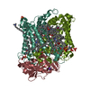
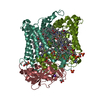
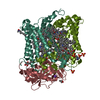
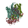
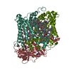
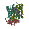
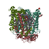
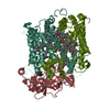
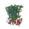
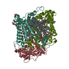
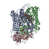
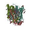
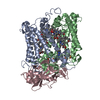
- PDB-2hhk: Reaction centre from Rhodobacter sphaeroides strain R-26.1 comple... -
+ Open data

- Basic information
| Entry | Database: PDB / ID: 2hhk | ||||||
|---|---|---|---|---|---|---|---|
| Title | Reaction centre from Rhodobacter sphaeroides strain R-26.1 complexed with dibrominated phosphatidylglycerol | ||||||
 Components Components | (Reaction center protein ...) x 3 | ||||||
 Keywords Keywords | PHOTOSYNTHESIS/MEMBRANE PROTEIN / PHOTOSYNTHESIS / PHOTOSYNTHETIC REACTION CENTER / LIPID BINDING SITES / BROMINATED LIPID / MEMBRANE PROTEIN / PHOTOSYNTHESIS-MEMBRANE PROTEIN COMPLEX | ||||||
| Function / homology |  Function and homology information Function and homology informationplasma membrane-derived chromatophore membrane / plasma membrane light-harvesting complex / bacteriochlorophyll binding / photosynthetic electron transport in photosystem II / photosynthesis, light reaction / metal ion binding Similarity search - Function | ||||||
| Biological species |  Rhodobacter sphaeroides (bacteria) Rhodobacter sphaeroides (bacteria) | ||||||
| Method |  X-RAY DIFFRACTION / SYNCHROTRON / MOLECULAR REPLACEMENT / Resolution: 2.5 Å X-RAY DIFFRACTION / SYNCHROTRON / MOLECULAR REPLACEMENT / Resolution: 2.5 Å | ||||||
 Authors Authors | Roszak, A.W. / Gardiner, A.T. / Isaacs, N.W. / Cogdell, R.J. | ||||||
 Citation Citation | Journal: Biochemistry / Year: 2007 Title: Brominated Lipids Identify Lipid Binding Sites on the Surface of the Reaction Center from Rhodobacter sphaeroides. Authors: Roszak, A.W. / Gardiner, A.T. / Isaacs, N.W. / Cogdell, R.J. | ||||||
| History |
|
- Structure visualization
| Structure viewer | Molecule: MolmilJmol/JSmol |
|---|
- Downloads & links
-Download
| PDBx/mmCIF format | 2hhk.cif.gz | 219.3 KB | Display | PDBx/mmCIF format |
|---|---|---|---|---|
| PDB format | pdb2hhk.ent.gz | 168 KB | Display | PDB format |
| PDBx/mmJSON format | 2hhk.json.gz | Tree view | PDBx/mmJSON format | |
| Others |  Other downloads Other downloads |
-Validation report
| Arichive directory | https://data.pdbj.org/pub/pdb/validation_reports/hh/2hhkftp://data.pdbj.org/pub/pdb/validation_reports/hh/2hhk | HTTPS FTP |
|---|
-Related structure data


-Links
 PDBj
PDBj


- Assembly
| Deposited unit | 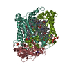
| ||||||||
|---|---|---|---|---|---|---|---|---|---|
| 1 |
| ||||||||
| Unit cell |
|
-Components
-Reaction center protein ... , 3 types, 3 molecules LMH
| #1: Protein | Mass: 31346.389 Da / Num. of mol.: 1 / Source method: isolated from a natural source Details: Strain R-26.1 of Rhodobacter sphaeroides bacteria is a partial revertant of the R-26 chemical mutant of the wild-type strain 2.4.1. While R-26 has no LH2 antenna and no carotenoid, the R-26. ...Details: Strain R-26.1 of Rhodobacter sphaeroides bacteria is a partial revertant of the R-26 chemical mutant of the wild-type strain 2.4.1. While R-26 has no LH2 antenna and no carotenoid, the R-26.1 has altered LH2 antenna and no carotenoid. Reaction center from R-26.1 strain is therefore identical with the wild-type strain 2.4.1 except for the missing carotenoid. Source: (natural) Rhodobacter sphaeroides (bacteria) / Strain: R26.1 / References: UniProt: P0C0Y8 |
|---|---|
| #2: Protein | Mass: 34398.543 Da / Num. of mol.: 1 / Source method: isolated from a natural source Details: Strain R-26.1 of Rhodobacter sphaeroides bacteria is a partial revertant of the R-26 chemical mutant of the wild-type strain 2.4.1. While R-26 has no LH2 antenna and no carotenoid, the R-26. ...Details: Strain R-26.1 of Rhodobacter sphaeroides bacteria is a partial revertant of the R-26 chemical mutant of the wild-type strain 2.4.1. While R-26 has no LH2 antenna and no carotenoid, the R-26.1 has altered LH2 antenna and no carotenoid. Reaction center from R-26.1 strain is therefore identical with the wild-type strain 2.4.1 except for the missing carotenoid. Source: (natural) Rhodobacter sphaeroides (bacteria) / Strain: R26.1 / References: UniProt: P0C0Y9 |
| #3: Protein | Mass: 28066.322 Da / Num. of mol.: 1 / Source method: isolated from a natural source Details: Strain R-26.1 of Rhodobacter sphaeroides bacteria is a partial revertant of the R-26 chemical mutant of the wild-type strain 2.4.1. While R-26 has no LH2 antenna and no carotenoid, the R-26. ...Details: Strain R-26.1 of Rhodobacter sphaeroides bacteria is a partial revertant of the R-26 chemical mutant of the wild-type strain 2.4.1. While R-26 has no LH2 antenna and no carotenoid, the R-26.1 has altered LH2 antenna and no carotenoid. Reaction center from R-26.1 strain is therefore identical with the wild-type strain 2.4.1 except for the missing carotenoid. Source: (natural) Rhodobacter sphaeroides (bacteria) / Strain: R26.1 / References: UniProt: P0C0Y7 |
-Non-polymers , 13 types, 438 molecules 


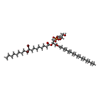


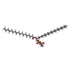

| #4: Chemical | ChemComp-BCL /  Mass: 911.504 Da / Num. of mol.: 4 / Source method: obtained synthetically / Formula: C55H74MgN4O6 Mass: 911.504 Da / Num. of mol.: 4 / Source method: obtained synthetically / Formula: C55H74MgN4O6#5: Chemical |  Mass: 889.215 Da / Num. of mol.: 2 / Source method: obtained synthetically / Formula: C55H76N4O6 Mass: 889.215 Da / Num. of mol.: 2 / Source method: obtained synthetically / Formula: C55H76N4O6#6: Chemical |  Mass: 863.343 Da / Num. of mol.: 2 / Source method: obtained synthetically / Formula: C59H90O4 Mass: 863.343 Da / Num. of mol.: 2 / Source method: obtained synthetically / Formula: C59H90O4#7: Chemical | ChemComp-GOL /  Mass: 92.094 Da / Num. of mol.: 4 / Source method: obtained synthetically / Formula: C3H8O3 Mass: 92.094 Da / Num. of mol.: 4 / Source method: obtained synthetically / Formula: C3H8O3#8: Chemical | ChemComp-FE / |  Mass: 55.845 Da / Num. of mol.: 1 / Source method: obtained synthetically / Formula: Fe Mass: 55.845 Da / Num. of mol.: 1 / Source method: obtained synthetically / Formula: Fe#9: Chemical | ChemComp-CL / |  Mass: 35.453 Da / Num. of mol.: 1 / Source method: obtained synthetically / Formula: Cl Mass: 35.453 Da / Num. of mol.: 1 / Source method: obtained synthetically / Formula: Cl#10: Chemical | ChemComp-PO4 /  Mass: 94.971 Da / Num. of mol.: 4 / Source method: obtained synthetically / Formula: PO4 Mass: 94.971 Da / Num. of mol.: 4 / Source method: obtained synthetically / Formula: PO4#11: Chemical | ChemComp-CDL / |  Mass: 1464.043 Da / Num. of mol.: 1 / Source method: obtained synthetically / Formula: C81H156O17P2 / Comment: phospholipid*YM Mass: 1464.043 Da / Num. of mol.: 1 / Source method: obtained synthetically / Formula: C81H156O17P2 / Comment: phospholipid*YM#12: Chemical | ChemComp-PGK / ( |  Mass: 908.815 Da / Num. of mol.: 1 / Source method: obtained synthetically / Formula: C40H77Br2O10P / Comment: phospholipid*YM Mass: 908.815 Da / Num. of mol.: 1 / Source method: obtained synthetically / Formula: C40H77Br2O10P / Comment: phospholipid*YM#13: Chemical | ChemComp-LDA /  Mass: 229.402 Da / Num. of mol.: 6 / Source method: obtained synthetically / Formula: C14H31NO / Comment: LDAO, detergent*YM Mass: 229.402 Da / Num. of mol.: 6 / Source method: obtained synthetically / Formula: C14H31NO / Comment: LDAO, detergent*YM#14: Chemical | ChemComp-K / |  Mass: 39.098 Da / Num. of mol.: 1 / Source method: obtained synthetically / Formula: K Mass: 39.098 Da / Num. of mol.: 1 / Source method: obtained synthetically / Formula: K#15: Chemical | ChemComp-PGT / ( |  Mass: 751.023 Da / Num. of mol.: 1 / Source method: obtained synthetically / Formula: C40H79O10P / Comment: phospholipid*YM Mass: 751.023 Da / Num. of mol.: 1 / Source method: obtained synthetically / Formula: C40H79O10P / Comment: phospholipid*YM#16: Water | ChemComp-HOH / | Mass: 18.015 Da / Num. of mol.: 410 / Source method: isolated from a natural source / Formula: H2O |
|---|
-Experimental details
-Experiment
| Experiment | Method: X-RAY DIFFRACTION / Number of used crystals: 1 |
|---|
- Sample preparation
| Crystal | Density Matthews: 5.49 Å3/Da / Density % sol: 77.59 % |
|---|---|
| Crystal grow | Temperature: 289 K / Method: vapor diffusion, sitting drop / pH: 8 Details: Potassium phosphate, LDAO, 1,2,3-heptanetriol, 1,2,3-hexanetriol, dioxane, NaCl, Tris-HCl, pH 8.0, VAPOR DIFFUSION, SITTING DROP, temperature 16.0K |
-Data collection
| Diffraction | Mean temperature: 100 K |
|---|---|
| Diffraction source | Source: SYNCHROTRON / Site: ESRF  / Beamline: BM14 / Wavelength: 0.9192 Å / Beamline: BM14 / Wavelength: 0.9192 Å |
| Detector | Type: MARMOSAIC 225 mm CCD / Detector: CCD / Date: Mar 18, 2005 / Details: mirrors |
| Radiation | Monochromator: Si(111) channel / Protocol: MAD / Monochromatic (M) / Laue (L): M / Scattering type: x-ray |
| Radiation wavelength | Wavelength: 0.9192 Å / Relative weight: 1 |
| Reflection | Resolution: 2.5→46.03 Å / Num. obs: 71485 / % possible obs: 99.6 % / Observed criterion σ(F): 0 / Observed criterion σ(I): 0 / Redundancy: 10.9 % / Biso Wilson estimate: 59.6 Å2 / Rmerge(I) obs: 0.059 / Net I/σ(I): 22.8 |
| Reflection shell | Resolution: 2.5→2.64 Å / Redundancy: 11 % / Rmerge(I) obs: 0.543 / Mean I/σ(I) obs: 4.6 / Num. unique all: 10292 / % possible all: 99.4 |
- Processing
| Software |
| |||||||||||||||||||||||||||||||||||||||||||||||||||||||||||||||||||||||||||||||||||||||||||||||||||||||||||||||||||||||||||||||||||||||||||||||||
|---|---|---|---|---|---|---|---|---|---|---|---|---|---|---|---|---|---|---|---|---|---|---|---|---|---|---|---|---|---|---|---|---|---|---|---|---|---|---|---|---|---|---|---|---|---|---|---|---|---|---|---|---|---|---|---|---|---|---|---|---|---|---|---|---|---|---|---|---|---|---|---|---|---|---|---|---|---|---|---|---|---|---|---|---|---|---|---|---|---|---|---|---|---|---|---|---|---|---|---|---|---|---|---|---|---|---|---|---|---|---|---|---|---|---|---|---|---|---|---|---|---|---|---|---|---|---|---|---|---|---|---|---|---|---|---|---|---|---|---|---|---|---|---|---|---|---|
| Refinement | Method to determine structure: MOLECULAR REPLACEMENT Starting model: Unpublished structure of reaction centre at 1.95A resolution Resolution: 2.5→46 Å / Cor.coef. Fo:Fc: 0.94 / Cor.coef. Fo:Fc free: 0.926 / SU B: 10.222 / SU ML: 0.104 / TLS residual ADP flag: LIKELY RESIDUAL Isotropic thermal model: TLS thermal mode followed by the restrained refinement of atomic coordinates and isotropic B-factors; all details in the pdb-file Cross valid method: THROUGHOUT / σ(F): 0 / σ(I): 0 / ESU R: 0.203 / ESU R Free: 0.17 / Stereochemistry target values: MAXIMUM LIKELIHOOD Details: HYDROGENS HAVE BEEN ADDED IN THE RIDING POSITIONS; the above average isotropic B value is an average residual B after TLS refinement while the final average atomic B is 64.8
| |||||||||||||||||||||||||||||||||||||||||||||||||||||||||||||||||||||||||||||||||||||||||||||||||||||||||||||||||||||||||||||||||||||||||||||||||
| Solvent computation | Ion probe radii: 0.8 Å / Shrinkage radii: 0.8 Å / VDW probe radii: 1.4 Å / Solvent model: BABINET MODEL WITH MASK | |||||||||||||||||||||||||||||||||||||||||||||||||||||||||||||||||||||||||||||||||||||||||||||||||||||||||||||||||||||||||||||||||||||||||||||||||
| Displacement parameters | Biso mean: 52.449 Å2
| |||||||||||||||||||||||||||||||||||||||||||||||||||||||||||||||||||||||||||||||||||||||||||||||||||||||||||||||||||||||||||||||||||||||||||||||||
| Refinement step | Cycle: LAST / Resolution: 2.5→46 Å
| |||||||||||||||||||||||||||||||||||||||||||||||||||||||||||||||||||||||||||||||||||||||||||||||||||||||||||||||||||||||||||||||||||||||||||||||||
| Refine LS restraints |
| |||||||||||||||||||||||||||||||||||||||||||||||||||||||||||||||||||||||||||||||||||||||||||||||||||||||||||||||||||||||||||||||||||||||||||||||||
| LS refinement shell | Resolution: 2.5→2.565 Å / Total num. of bins used: 20
| |||||||||||||||||||||||||||||||||||||||||||||||||||||||||||||||||||||||||||||||||||||||||||||||||||||||||||||||||||||||||||||||||||||||||||||||||
| Refinement TLS params. | Method: refined / Origin x: 55.9552 Å / Origin y: 60.7283 Å / Origin z: 64.6778 Å
| |||||||||||||||||||||||||||||||||||||||||||||||||||||||||||||||||||||||||||||||||||||||||||||||||||||||||||||||||||||||||||||||||||||||||||||||||
| Refinement TLS group |
|
