 Movie
Movie Controller

[English] 日本語
 Yorodumi
- PDB-2hex: DECAMERS OBSERVED IN THE CRYSTALS OF BOVINE PANCREATIC TRYPSIN IN... -
+ Open data

- Basic information
| Entry | Database: PDB / ID: 2hex | ||||||
|---|---|---|---|---|---|---|---|
| Title | DECAMERS OBSERVED IN THE CRYSTALS OF BOVINE PANCREATIC TRYPSIN INHIBITOR | ||||||
 Components Components | PROTEIN (PANCREATIC TRYPSIN INHIBITOR) | ||||||
 Keywords Keywords | BLOOD CLOTTING / BOVINE PANCREATIC TRYPSIN INHIBITOR / PENTAMERIC MOLECULE | ||||||
| Function / homology |  Function and homology information Function and homology informationsulfate binding / negative regulation of platelet aggregation / zymogen binding / potassium channel inhibitor activity / molecular function inhibitor activity / negative regulation of thrombin-activated receptor signaling pathway / serine protease inhibitor complex / serine-type endopeptidase inhibitor activity / protease binding / calcium ion binding / : Similarity search - Function | ||||||
| Biological species |  | ||||||
| Method |  X-RAY DIFFRACTION / SYNCHROTRON / MOLECULAR REPLACEMENT / Resolution: 2.1 Å X-RAY DIFFRACTION / SYNCHROTRON / MOLECULAR REPLACEMENT / Resolution: 2.1 Å | ||||||
 Authors Authors | Lubkowski, J. / Wlodawer, A. | ||||||
 Citation Citation | Journal: Acta Crystallogr.,Sect.D / Year: 1999 Title: Decamers observed in the crystals of bovine pancreatic trypsin inhibitor. Authors: Lubkowski, J. / Wlodawer, A. | ||||||
| History |
|
- Structure visualization
| Structure viewer | Molecule: MolmilJmol/JSmol |
|---|
- Downloads & links
-Download
| PDBx/mmCIF format | 2hex.cif.gz | 72 KB | Display | PDBx/mmCIF format |
|---|---|---|---|---|
| PDB format | pdb2hex.ent.gz | 55.1 KB | Display | PDB format |
| PDBx/mmJSON format | 2hex.json.gz | Tree view | PDBx/mmJSON format | |
| Others |  Other downloads Other downloads |
-Validation report
| Arichive directory | https://data.pdbj.org/pub/pdb/validation_reports/he/2hexftp://data.pdbj.org/pub/pdb/validation_reports/he/2hex | HTTPS FTP |
|---|
-Related structure data
| Related structure data |  1bpiS S: Starting model for refinement |
|---|---|
| Similar structure data |


-Links
 PDBj
PDBj


- Assembly
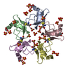
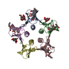
| Deposited unit | 
| ||||||||
|---|---|---|---|---|---|---|---|---|---|
| 1 |
| ||||||||
| 2 | 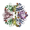
| ||||||||
| Unit cell |
| ||||||||
| Components on special symmetry positions |
|
-Components
| #1: Protein | Mass: 6527.568 Da / Num. of mol.: 5 / Source method: isolated from a natural source / Source: (natural) #2: Chemical | ChemComp-SO4 /   Mass: 96.063 Da / Num. of mol.: 8 / Source method: obtained synthetically / Formula: SO4 Mass: 96.063 Da / Num. of mol.: 8 / Source method: obtained synthetically / Formula: SO4#3: Water | ChemComp-HOH / |  Mass: 18.015 Da / Num. of mol.: 181 / Source method: isolated from a natural source / Formula: H2O Mass: 18.015 Da / Num. of mol.: 181 / Source method: isolated from a natural source / Formula: H2OHas protein modification | Y | |
|---|
-Experimental details
-Experiment
| Experiment | Method: X-RAY DIFFRACTION / Number of used crystals: 1 |
|---|
- Sample preparation
| Crystal | Density Matthews: 2.34 Å3/Da / Density % sol: 47.4 % | ||||||||||||||||||||
|---|---|---|---|---|---|---|---|---|---|---|---|---|---|---|---|---|---|---|---|---|---|
| Crystal grow | Method: vapor diffusion, hanging drop / pH: 6.5 Details: USING SOLUTION #32 IN CRYSTAL SCREEN I (HAMPTON RESEARCH), pH 6.5, VAPOR DIFFUSION, HANGING DROP | ||||||||||||||||||||
| Crystal | *PLUS | ||||||||||||||||||||
| Crystal grow | *PLUS pH: 7 / Method: vapor diffusion, sitting drop | ||||||||||||||||||||
| Components of the solutions | *PLUS
|
-Data collection
| Diffraction | Mean temperature: 100 K |
|---|---|
| Diffraction source | Source: SYNCHROTRON / Site: NSLS  / Beamline: X9B / Wavelength: 0.99 / Beamline: X9B / Wavelength: 0.99 |
| Detector | Type: MARRESEARCH / Detector: IMAGE PLATE / Date: Feb 15, 1997 / Details: MIRRORS |
| Radiation | Monochromator: SILICON CRYSTAL / Protocol: SINGLE WAVELENGTH / Monochromatic (M) / Laue (L): M / Scattering type: x-ray |
| Radiation wavelength | Wavelength: 0.99 Å / Relative weight: 1 |
| Reflection | Resolution: 2.1→20 Å / Num. obs: 25235 / % possible obs: 99.6 % / Observed criterion σ(I): -3 / Redundancy: 6.82 % / Rmerge(I) obs: 0.074 / Rsym value: 0.074 / Net I/σ(I): 23 |
| Reflection shell | Resolution: 2.1→2.17 Å / Rmerge(I) obs: 0.47 / Mean I/σ(I) obs: 4.3 / Rsym value: 0.47 / % possible all: 99 |
| Reflection | *PLUS Num. measured all: 333309 / Rmerge(I) obs: 0.074 |
| Reflection shell | *PLUS % possible obs: 99 % |
- Processing
| Software |
| |||||||||||||||||||||||||||||||||
|---|---|---|---|---|---|---|---|---|---|---|---|---|---|---|---|---|---|---|---|---|---|---|---|---|---|---|---|---|---|---|---|---|---|---|
| Refinement | Method to determine structure: MOLECULAR REPLACEMENT Starting model: 1BPI Resolution: 2.1→10 Å / Num. parameters: 9897 / Num. restraintsaints: 9448 / Cross valid method: FREE R / σ(F): 0 / Stereochemistry target values: ENGH AND HUBER Details: ANISOTROPIC SCALING APPLIED BY THE METHOD OF PARKIN, MOEZZI & HOPE, J.APPL.CRYST.28 (1995)53-56 B23 (A**2) : ESTIMATED OVERALL COORDINATE ERROR.
| |||||||||||||||||||||||||||||||||
| Solvent computation | Solvent model: MOEWS & KRETSINGER, J.MOL.BIOL.91(1973)201-228 | |||||||||||||||||||||||||||||||||
| Refine analyze | Num. disordered residues: 3 / Occupancy sum hydrogen: 0 / Occupancy sum non hydrogen: 2446.5 | |||||||||||||||||||||||||||||||||
| Refinement step | Cycle: LAST / Resolution: 2.1→10 Å
| |||||||||||||||||||||||||||||||||
| Refine LS restraints |
| |||||||||||||||||||||||||||||||||
| Software | *PLUS Name: SHELXL-97 / Classification: refinement | |||||||||||||||||||||||||||||||||
| Refinement | *PLUS Lowest resolution: 10 Å / σ(F): 0 / % reflection Rfree: 11.2 % / Rfactor Rwork: 0.207 | |||||||||||||||||||||||||||||||||
| Solvent computation | *PLUS | |||||||||||||||||||||||||||||||||
| Displacement parameters | *PLUS | |||||||||||||||||||||||||||||||||
| Refine LS restraints | *PLUS Type: s_plane_restr / Dev ideal: 0.025 |
