 Movie
Movie Controller

[English] 日本語
 Yorodumi
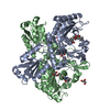
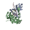
- PDB-1b0c: EVIDENCE OF A COMMON DECAMER IN THREE CRYSTAL STRUCTURES OF BPTI,... -
+ Open data

- Basic information
| Entry | Database: PDB / ID: 1b0c | ||||||
|---|---|---|---|---|---|---|---|
| Title | EVIDENCE OF A COMMON DECAMER IN THREE CRYSTAL STRUCTURES OF BPTI, CRYSTALLIZED FROM THIOCYANATE, CHLORIDE OR SULFATE | ||||||
 Components Components | PROTEIN (PANCREATIC TRYPSIN INHIBITOR) | ||||||
 Keywords Keywords | HYDROLASE INHIBITOR / BOVINE PANCREATIC TRYPSIN INHIBITOR / PENTAMERIC MOLECULE | ||||||
| Function / homology |  Function and homology information Function and homology informationsulfate binding / negative regulation of platelet aggregation / zymogen binding / potassium channel inhibitor activity / molecular function inhibitor activity / negative regulation of thrombin-activated receptor signaling pathway / serine protease inhibitor complex / serine-type endopeptidase inhibitor activity / protease binding / calcium ion binding / : Similarity search - Function | ||||||
| Biological species |  | ||||||
| Method |  X-RAY DIFFRACTION / SYNCHROTRON / MOLECULAR REPLACEMENT / Resolution: 2.8 Å X-RAY DIFFRACTION / SYNCHROTRON / MOLECULAR REPLACEMENT / Resolution: 2.8 Å | ||||||
 Authors Authors | Hamiaux, C. / Prange, T. / Ries-Kautt, M. / Ducruix, A. / Lafont, S. / Astier, J.P. / Veesler, S. | ||||||
 Citation Citation | Journal: J.Mol.Biol. / Year: 2000 Title: The BPTI decamer observed in acidic pH crystal forms pre-exists as a stable species in solution. Authors: Hamiaux, C. / Perez, J. / Prange, T. / Veesler, S. / Ries-Kautt, M. / Vachette, P. #1: Journal: Acta Crystallogr.,Sect.D / Year: 1999Title: The Decameric Structure of Bovine Pancreatic Trypsin Inhibitor (Bpti) Crystallized from Thiocyanate at 2.7A Resolution Authors: Hamiaux, C. / Prange, T. / Ries-Kautt, M. / Ducruix, A. / Lafont, S. / Astier, J.P. / Veesler, S. #2: Journal: Acta Crystallogr.,Sect.D / Year: 1999Title: Decamers Observed in the Crystals of Bovine Panreatic Trypsin Inhibitor Authors: Lubkowski, J. / Wlodawer, A. | ||||||
| History |
|
- Structure visualization
| Structure viewer | Molecule: MolmilJmol/JSmol |
|---|
- Downloads & links
-Download
| PDBx/mmCIF format | 1b0c.cif.gz | 60.9 KB | Display | PDBx/mmCIF format |
|---|---|---|---|---|
| PDB format | pdb1b0c.ent.gz | 46 KB | Display | PDB format |
| PDBx/mmJSON format | 1b0c.json.gz | Tree view | PDBx/mmJSON format | |
| Others |  Other downloads Other downloads |
-Validation report
| Arichive directory | https://data.pdbj.org/pub/pdb/validation_reports/b0/1b0cftp://data.pdbj.org/pub/pdb/validation_reports/b0/1b0c | HTTPS FTP |
|---|
-Related structure data
| Related structure data |  1bz5C 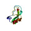 6ptiS S: Starting model for refinement C: citing same article ( |
|---|---|
| Similar structure data |

-Links
 PDBj
PDBj


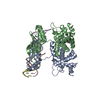
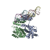
- Assembly
| Deposited unit | 
| |||||||||||||||||||||||||||||||||||
|---|---|---|---|---|---|---|---|---|---|---|---|---|---|---|---|---|---|---|---|---|---|---|---|---|---|---|---|---|---|---|---|---|---|---|---|---|
| 1 | 
| |||||||||||||||||||||||||||||||||||
| Unit cell |
| |||||||||||||||||||||||||||||||||||
| Noncrystallographic symmetry (NCS) | NCS domain:
NCS oper:
|
-Components
| #1: Protein | Mass: 6527.568 Da / Num. of mol.: 5 Source method: isolated from a genetically manipulated source Source: (gene. exp.) #2: Water | ChemComp-HOH / |  Mass: 18.015 Da / Num. of mol.: 58 / Source method: isolated from a natural source / Formula: H2O Mass: 18.015 Da / Num. of mol.: 58 / Source method: isolated from a natural source / Formula: H2OHas protein modification | Y | |
|---|
-Experimental details
-Experiment
| Experiment | Method: X-RAY DIFFRACTION / Number of used crystals: 1 |
|---|
- Sample preparation
| Crystal | Density Matthews: 3.23 Å3/Da / Density % sol: 61 % | ||||||||||||||||||||||||
|---|---|---|---|---|---|---|---|---|---|---|---|---|---|---|---|---|---|---|---|---|---|---|---|---|---|
| Crystal grow | pH: 4.5 Details: ACETATE BUFFER PH=4.5 BPTI 60 TO 100 MG/ML NACL 1.6 TO 2 M | ||||||||||||||||||||||||
| Crystal | *PLUS | ||||||||||||||||||||||||
| Crystal grow | *PLUS Temperature: 18 ℃ / Method: vapor diffusion, hanging drop | ||||||||||||||||||||||||
| Components of the solutions | *PLUS
|
-Data collection
| Diffraction | Mean temperature: 297 K |
|---|---|
| Diffraction source | Source: SYNCHROTRON / Site: LURE  / Beamline: DW32 / Wavelength: 0.97 / Beamline: DW32 / Wavelength: 0.97 |
| Detector | Type: MAR scanner 345 mm plate / Detector: IMAGE PLATE / Date: Mar 15, 1997 / Details: MIRRORS |
| Radiation | Monochromator: SILICON CRYSTAL / Protocol: SINGLE WAVELENGTH / Monochromatic (M) / Laue (L): M / Scattering type: x-ray |
| Radiation wavelength | Wavelength: 0.97 Å / Relative weight: 1 |
| Reflection | Resolution: 2.8→18 Å / Num. obs: 11127 / % possible obs: 99.6 % / Observed criterion σ(I): 2 / Redundancy: 9.6 % / Biso Wilson estimate: 55.8 Å2 / Rsym value: 0.038 / Net I/σ(I): 17.2 |
| Reflection shell | Resolution: 2.8→2.87 Å / Redundancy: 9.8 % / Mean I/σ(I) obs: 5.4 / Rsym value: 0.14 / % possible all: 99 |
| Reflection | *PLUS Highest resolution: 2.77 Å / % possible obs: 89.3 % / Num. measured all: 107368 / Rmerge(I) obs: 0.038 |
| Reflection shell | *PLUS Highest resolution: 2.77 Å / Lowest resolution: 2.84 Å / % possible obs: 55.5 % / Rmerge(I) obs: 0.161 / Mean I/σ(I) obs: 4.7 |
- Processing
| Software |
| ||||||||||||||||||||||||||||||||||||||||||||||||||||||||||||
|---|---|---|---|---|---|---|---|---|---|---|---|---|---|---|---|---|---|---|---|---|---|---|---|---|---|---|---|---|---|---|---|---|---|---|---|---|---|---|---|---|---|---|---|---|---|---|---|---|---|---|---|---|---|---|---|---|---|---|---|---|---|
| Refinement | Method to determine structure: MOLECULAR REPLACEMENT Starting model: 6PTI Resolution: 2.8→18 Å / Rfactor Rfree error: 0.007 / Data cutoff high absF: 10000000 / Data cutoff low absF: 0.001 / Isotropic thermal model: GROUP / Cross valid method: THROUGHOUT / σ(F): 2 / Details: BULK SOLVENT MODEL USED
| ||||||||||||||||||||||||||||||||||||||||||||||||||||||||||||
| Displacement parameters | Biso mean: 37.1 Å2
| ||||||||||||||||||||||||||||||||||||||||||||||||||||||||||||
| Refine analyze | Luzzati coordinate error obs: 0.35 Å / Luzzati d res low obs: 5 Å / Luzzati sigma a obs: 0.38 Å | ||||||||||||||||||||||||||||||||||||||||||||||||||||||||||||
| Refinement step | Cycle: LAST / Resolution: 2.8→18 Å
| ||||||||||||||||||||||||||||||||||||||||||||||||||||||||||||
| Refine LS restraints |
| ||||||||||||||||||||||||||||||||||||||||||||||||||||||||||||
| Refine LS restraints NCS |
| ||||||||||||||||||||||||||||||||||||||||||||||||||||||||||||
| LS refinement shell | Resolution: 2.8→2.9 Å / Rfactor Rfree error: 0.033 / Total num. of bins used: 10
| ||||||||||||||||||||||||||||||||||||||||||||||||||||||||||||
| Xplor file |
| ||||||||||||||||||||||||||||||||||||||||||||||||||||||||||||
| Software | *PLUS Name: X-PLOR / Version: 3.851 / Classification: refinement | ||||||||||||||||||||||||||||||||||||||||||||||||||||||||||||
| Refinement | *PLUS σ(F): 2 / % reflection Rfree: 10.3 % | ||||||||||||||||||||||||||||||||||||||||||||||||||||||||||||
| Solvent computation | *PLUS | ||||||||||||||||||||||||||||||||||||||||||||||||||||||||||||
| Displacement parameters | *PLUS Biso mean: 37.1 Å2 | ||||||||||||||||||||||||||||||||||||||||||||||||||||||||||||
| Refine LS restraints | *PLUS
| ||||||||||||||||||||||||||||||||||||||||||||||||||||||||||||
| LS refinement shell | *PLUS Rfactor Rfree: 0.348 / % reflection Rfree: 10.2 % / Rfactor Rwork: 0.3 |
