SEQUENCE THE CONSTRUCT WAS EXPRESSED WITH A PURIFICATION TAG MGSDKIHHHHHHENLYFQG. THE TAG WAS ...SEQUENCE THE CONSTRUCT WAS EXPRESSED WITH A PURIFICATION TAG MGSDKIHHHHHHENLYFQG. THE TAG WAS REMOVED WITH TEV PROTEASE LEAVING ONLY A GLYCINE (0) FOLLOWED BY THE TARGET SEQUENCE.
Monochromator: Single crystal Si(111) bent monochromator (horizontal focusing) Protocol: MAD / Monochromatic (M) / Laue (L): M / Scattering type: x-ray
Radiation wavelength
ID
Wavelength (Å)
Relative weight
1
0.979224
1
2
0.978954
1
3
0.91837
1
Reflection
Resolution: 1.5→29.424 Å / Num. obs: 32658 / % possible obs: 96.5 % / Redundancy: 8.784 % / Biso Wilson estimate: 22.126 Å2 / Rmerge(I) obs: 0.143 / Net I/σ(I): 7.33
Reflection shell
Diffraction-ID: 1
Resolution (Å)
Highest resolution (Å)
Rmerge(I) obs
Mean I/σ(I) obs
Num. measured obs
Num. unique all
% possible all
1.5-1.55
0.426
2.32
11737
4246
88.8
1.55-1.62
0.382
2.9
18629
6449
89
1.62-1.69
0.317
3.5
15873
5490
90.6
1.69-1.78
0.447
4.4
23105
5918
91.5
1.78-1.89
0.468
5.3
34402
5928
93.2
1.89-2.04
0.356
6.6
33794
5949
91.3
2.04-2.24
0.237
8.1
34138
6057
96.4
2.24-2.56
0.163
9.9
34882
6205
97.6
2.56
0.113
12.8
36393
6372
98.2
-
Phasing
Phasing
Method: MAD
-
Processing
Software
Name
Version
Classification
NB
MolProbity
3beta29
modelbuilding
REFMAC
5.2.0005
refinement
XSCALE
datascaling
PDB_EXTRACT
2
dataextraction
XDS
datareduction
Refinement
Method to determine structure: MAD / Resolution: 1.5→29.424 Å / Cor.coef. Fo:Fc: 0.966 / Cor.coef. Fo:Fc free: 0.954 / SU B: 3.408 / SU ML: 0.062 / TLS residual ADP flag: LIKELY RESIDUAL / Cross valid method: THROUGHOUT / σ(F): 0 / ESU R: 0.079 / ESU R Free: 0.081 Stereochemistry target values: MAXIMUM LIKELIHOOD WITH PHASES Details: 1.HYDROGENS HAVE BEEN ADDED IN THE RIDING POSITIONS. 2.A MET-INHIBITION PROTOCOL WAS USED FOR SELENOMETHIONINE INCORPORATION DURING PROTEIN EXPRESSION. THE OCCUPANCY OF THE SE ATOMS IN THE ...Details: 1.HYDROGENS HAVE BEEN ADDED IN THE RIDING POSITIONS. 2.A MET-INHIBITION PROTOCOL WAS USED FOR SELENOMETHIONINE INCORPORATION DURING PROTEIN EXPRESSION. THE OCCUPANCY OF THE SE ATOMS IN THE MSE RESIDUES WAS REDUCED TO 0.75 TO ACCOUNT FOR THE REDUCED SCATTERING POWER DUE TO PARTIAL S-MET INCORPORATION. 3.DUE TO SEVERAL STRONG ICE RINGS, 1316 REFLECTIONS WITH INTENSITIES >=15 * (EXPECTED MEAN INTENSITY) BETWEEN 1.516-1.533, 1.890-1.930, 2.025-2.080, 2.210-2.289 ANGSTROMS WERE OMITTED FROM THE FINAL REFINEMENT. 4.TWO PHOSPHATE IONS FROM CRYSTALLIZATION BUFFER WERE MODELED.
Rfactor
Num. reflection
% reflection
Selection details
Rfree
0.209
1564
5.1 %
RANDOM
Rwork
0.176
-
-
-
obs
0.178
30840
95.21 %
-
Solvent computation
Ion probe radii: 0.8 Å / Shrinkage radii: 0.8 Å / VDW probe radii: 1.2 Å / Solvent model: MASK
Displacement parameters
Biso mean: 15.763 Å2
Baniso -1
Baniso -2
Baniso -3
1-
-0.65 Å2
-0.33 Å2
0 Å2
2-
-
-0.65 Å2
0 Å2
3-
-
-
0.98 Å2
Refinement step
Cycle: LAST / Resolution: 1.5→29.424 Å
Protein
Nucleic acid
Ligand
Solvent
Total
Num. atoms
1574
0
10
212
1796
Refine LS restraints
Refine-ID
Type
Dev ideal
Dev ideal target
Number
X-RAY DIFFRACTION
r_bond_refined_d
0.014
0.022
1665
X-RAY DIFFRACTION
r_bond_other_d
0.001
0.02
1489
X-RAY DIFFRACTION
r_angle_refined_deg
1.478
1.964
2278
X-RAY DIFFRACTION
r_angle_other_deg
0.808
3
3468
X-RAY DIFFRACTION
r_dihedral_angle_1_deg
5.575
5
220
X-RAY DIFFRACTION
r_dihedral_angle_2_deg
35.027
24.714
70
X-RAY DIFFRACTION
r_dihedral_angle_3_deg
12.157
15
265
X-RAY DIFFRACTION
r_dihedral_angle_4_deg
14.608
15
7
X-RAY DIFFRACTION
r_chiral_restr
0.084
0.2
257
X-RAY DIFFRACTION
r_gen_planes_refined
0.006
0.02
1881
X-RAY DIFFRACTION
r_gen_planes_other
0.001
0.02
325
X-RAY DIFFRACTION
r_nbd_refined
0.229
0.2
377
X-RAY DIFFRACTION
r_nbd_other
0.173
0.2
1473
X-RAY DIFFRACTION
r_nbtor_refined
0.183
0.2
835
X-RAY DIFFRACTION
r_nbtor_other
0.086
0.2
940
X-RAY DIFFRACTION
r_xyhbond_nbd_refined
0.144
0.2
142
X-RAY DIFFRACTION
r_symmetry_vdw_refined
0.088
0.2
5
X-RAY DIFFRACTION
r_symmetry_vdw_other
0.12
0.2
31
X-RAY DIFFRACTION
r_symmetry_hbond_refined
0.163
0.2
12
X-RAY DIFFRACTION
r_mcbond_it
1.984
3
1093
X-RAY DIFFRACTION
r_mcbond_other
0.486
3
420
X-RAY DIFFRACTION
r_mcangle_it
2.765
5
1727
X-RAY DIFFRACTION
r_scbond_it
4.578
8
643
X-RAY DIFFRACTION
r_scangle_it
6.504
11
546
LS refinement shell
Resolution: 1.504→1.543 Å / Total num. of bins used: 20
Rfactor
Num. reflection
% reflection
Rfree
0.368
84
-
Rwork
0.295
1597
-
obs
-
1681
70.31 %
Refinement TLS params.
Method: refined / Origin x: 24.3027 Å / Origin y: 21.9784 Å / Origin z: 22.2181 Å
11
12
13
21
22
23
31
32
33
T
-0.0187 Å2
0.0106 Å2
0.0093 Å2
-
-0.0232 Å2
0.0104 Å2
-
-
-0.0273 Å2
L
0.6919 °2
-0.1209 °2
0.1494 °2
-
0.7872 °2
0.5101 °2
-
-
0.7208 °2
S
-0.0157 Å °
0.0103 Å °
0.0008 Å °
0.0465 Å °
0.0182 Å °
0.0121 Å °
0.0598 Å °
0.0418 Å °
-0.0025 Å °
Refinement TLS group
Selection: ALL
+
About Yorodumi
-
News
-
Feb 9, 2022. New format data for meta-information of EMDB entries
New format data for meta-information of EMDB entries
Version 3 of the EMDB header file is now the official format.
The previous official version 1.9 will be removed from the archive.
In the structure databanks used in Yorodumi, some data are registered as the other names, "COVID-19 virus" and "2019-nCoV". Here are the details of the virus and the list of structure data.
Jan 31, 2019. EMDB accession codes are about to change! (news from PDBe EMDB page)
EMDB accession codes are about to change! (news from PDBe EMDB page)
The allocation of 4 digits for EMDB accession codes will soon come to an end. Whilst these codes will remain in use, new EMDB accession codes will include an additional digit and will expand incrementally as the available range of codes is exhausted. The current 4-digit format prefixed with “EMD-” (i.e. EMD-XXXX) will advance to a 5-digit format (i.e. EMD-XXXXX), and so on. It is currently estimated that the 4-digit codes will be depleted around Spring 2019, at which point the 5-digit format will come into force.
The EM Navigator/Yorodumi systems omit the EMD- prefix.
Related info.:Q: What is EMD? / ID/Accession-code notation in Yorodumi/EM Navigator
Yorodumi is a browser for structure data from EMDB, PDB, SASBDB, etc.
This page is also the successor to EM Navigator detail page, and also detail information page/front-end page for Omokage search.
The word "yorodu" (or yorozu) is an old Japanese word meaning "ten thousand". "mi" (miru) is to see.
Related info.:EMDB / PDB / SASBDB / Comparison of 3 databanks / Yorodumi Search / Aug 31, 2016. New EM Navigator & Yorodumi / Yorodumi Papers / Jmol/JSmol / Function and homology information / Changes in new EM Navigator and Yorodumi
 Movie
Movie Controller

 Yorodumi
 Open data
 Basic information
 Components
 Keywords
 Function and homology information
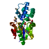
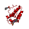
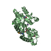
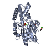
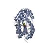
 Lactobacillus plantarum (bacteria)
 X-RAY DIFFRACTION /
  Authors
 Citation
 Structure visualization
 Downloads & links
 Other downloads


 PDBj
PDBj
 Assembly

 Mass: 94.971 Da / Num. of mol.: 2 / Source method: obtained synthetically / Formula: PO4
 Mass: 18.015 Da / Num. of mol.: 212 / Source method: isolated from a natural source / Formula: H2O
 Sample preparation
 / Beamline: BL11-1 / Wavelength: 0.979224, 0.978954, 0.918370
 Processing