Mass: 18.015 Da / Num. of mol.: 176 / Source method: isolated from a natural source / Formula: H2O
-
Experimental details
-
Experiment
Experiment
Method: X-RAY DIFFRACTION / Number of used crystals: 1
-
Sample preparation
Crystal
Density Matthews: 2.72 Å3/Da / Density % sol: 54.8 %
Crystal grow
Temperature: 277 K / Method: vapor diffusion, hanging drop / pH: 8.5 Details: 0.1 M Tris pH 8.5 0.8 M Ammonium Acetate 0.25 M Lithium Chloride 12% PEG 3350, VAPOR DIFFUSION, HANGING DROP, temperature 277.0K
-
Data collection
Diffraction
ID
Mean temperature (K)
Crystal-ID
1
93
1
2
93
1
3
93
1
Diffraction source
Source
Site
Beamline
ID
Wavelength (Å)
SYNCHROTRON
NSLS
X9A
1
0.97927
SYNCHROTRON
NSLS
X9A
2
0.97939
SYNCHROTRON
NSLS
X29A
3
1.1
Detector
Type
ID
Detector
Date
MAR CCD 165 mm
1
CCD
Nov 18, 2004
MAR CCD 165 mm
2
CCD
Nov 18, 2004
ADSC QUANTUM 315
3
CCD
Mar 2, 2005
Radiation
ID
Monochromator
Protocol
Monochromatic (M) / Laue (L)
Scattering type
Wavelength-ID
1
Si111
MAD
M
x-ray
1
2
Si111
MAD
M
x-ray
1
3
Si111
SINGLEWAVELENGTH
M
x-ray
1
Radiation wavelength
ID
Wavelength (Å)
Relative weight
1
0.97927
1
2
0.97939
1
3
1.1
1
Reflection
Resolution: 2.28→30 Å / Num. obs: 75215 / % possible obs: 99.6 % / Observed criterion σ(I): 4 / Redundancy: 3.4 % / Rsym value: 0.087 / Net I/σ(I): 13.1
Reflection shell
Resolution: 2.28→2.36 Å / Redundancy: 3.2 % / Rsym value: 0.175 / % possible all: 96.4
-
Processing
Software
Name
Version
Classification
MOLREP
phasing
CNS
1.1
refinement
DENZO
datareduction
HKL-2000
datascaling
Refinement
Method to determine structure: MOLECULAR REPLACEMENT / Resolution: 2.28→30 Å / σ(F): 2 / Stereochemistry target values: Engh & Huber
Rfactor
Num. reflection
Selection details
Rfree
0.268
7521
RANDOM
Rwork
0.226
-
-
all
-
82381
-
obs
-
75215
-
Refinement step
Cycle: LAST / Resolution: 2.28→30 Å
Protein
Nucleic acid
Ligand
Solvent
Total
Num. atoms
11501
0
253
176
11930
Refine LS restraints
Refine-ID
Type
Dev ideal
X-RAY DIFFRACTION
c_bond_d
0.007
X-RAY DIFFRACTION
c_angle_deg
1.3
+
About Yorodumi
-
News
-
Feb 9, 2022. New format data for meta-information of EMDB entries
New format data for meta-information of EMDB entries
Version 3 of the EMDB header file is now the official format.
The previous official version 1.9 will be removed from the archive.
In the structure databanks used in Yorodumi, some data are registered as the other names, "COVID-19 virus" and "2019-nCoV". Here are the details of the virus and the list of structure data.
Jan 31, 2019. EMDB accession codes are about to change! (news from PDBe EMDB page)
EMDB accession codes are about to change! (news from PDBe EMDB page)
The allocation of 4 digits for EMDB accession codes will soon come to an end. Whilst these codes will remain in use, new EMDB accession codes will include an additional digit and will expand incrementally as the available range of codes is exhausted. The current 4-digit format prefixed with “EMD-” (i.e. EMD-XXXX) will advance to a 5-digit format (i.e. EMD-XXXXX), and so on. It is currently estimated that the 4-digit codes will be depleted around Spring 2019, at which point the 5-digit format will come into force.
The EM Navigator/Yorodumi systems omit the EMD- prefix.
Related info.:Q: What is EMD? / ID/Accession-code notation in Yorodumi/EM Navigator
Yorodumi is a browser for structure data from EMDB, PDB, SASBDB, etc.
This page is also the successor to EM Navigator detail page, and also detail information page/front-end page for Omokage search.
The word "yorodu" (or yorozu) is an old Japanese word meaning "ten thousand". "mi" (miru) is to see.
Related info.:EMDB / PDB / SASBDB / Comparison of 3 databanks / Yorodumi Search / Aug 31, 2016. New EM Navigator & Yorodumi / Yorodumi Papers / Jmol/JSmol / Function and homology information / Changes in new EM Navigator and Yorodumi
 Movie
Movie Controller


 Open data
 Basic information
 Components
 Keywords
 Function and homology information
 Hepatitis C virus
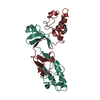
 X-RAY DIFFRACTION /
  Authors
 Citation
 Structure visualization
 Downloads & links
 Other downloads


 PDBj
PDBj

 Assembly


 Type: D-saccharide / Mass: 292.369 Da / Num. of mol.: 11
 Type: D-saccharide / Mass: 482.562 Da / Num. of mol.: 1
 Mass: 18.015 Da / Num. of mol.: 176 / Source method: isolated from a natural source / Formula: H2O
 Sample preparation
 Processing