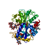
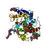
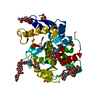
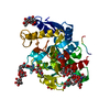
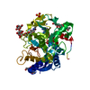
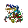
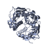
Metal sequestration by antimicrobial proteins / Antimicrobial peptides / negative regulation of tumor necrosis factor (ligand) superfamily member 11 production / negative regulation of single-species biofilm formation in or on host organism / positive regulation of bone mineralization involved in bone maturation / negative regulation of osteoclast development / specific granule / negative regulation of lipopolysaccharide-mediated signaling pathway / positive regulation of chondrocyte proliferation / antifungal humoral response ...Metal sequestration by antimicrobial proteins / Antimicrobial peptides / negative regulation of tumor necrosis factor (ligand) superfamily member 11 production / negative regulation of single-species biofilm formation in or on host organism / positive regulation of bone mineralization involved in bone maturation / negative regulation of osteoclast development / specific granule / negative regulation of lipopolysaccharide-mediated signaling pathway / positive regulation of chondrocyte proliferation / antifungal humoral response / regulation of tumor necrosis factor production / bone morphogenesis / Neutrophil degranulation / positive regulation of osteoblast proliferation / Hydrolases; Acting on peptide bonds (peptidases); Serine endopeptidases / positive regulation of osteoblast differentiation / cysteine-type endopeptidase inhibitor activity / regulation of cytokine production / ossification / innate immune response in mucosa / iron ion transport / recycling endosome / antibacterial humoral response / early endosome / iron ion binding / signaling receptor binding / serine-type endopeptidase activity / negative regulation of apoptotic process / proteolysis / : / plasma membrane Similarity search - Function
Resolution: 2.8→20 Å / Cor.coef. Fo:Fc: 0.907 / Cor.coef. Fo:Fc free: 0.825 / SU B: 14.897 / SU ML: 0.306 / Cross valid method: THROUGHOUT / σ(F): 0 / ESU R Free: 0.475 / Stereochemistry target values: MAXIMUM LIKELIHOOD / Details: HYDROGENS HAVE BEEN ADDED IN THE RIDING POSITIONS
Rfactor
Num. reflection
% reflection
Selection details
Rfree
0.24833
455
4.8 %
RANDOM
Rwork
0.21437
-
-
-
all
0.21272
9101
-
-
obs
0.21812
9081
96.32 %
-
Solvent computation
Ion probe radii: 0.8 Å / Shrinkage radii: 0.8 Å / VDW probe radii: 1.4 Å / Solvent model: MASK
Displacement parameters
Biso mean: 30.288 Å2
Baniso -1
Baniso -2
Baniso -3
1-
1.54 Å2
0 Å2
-0.08 Å2
2-
-
-0.75 Å2
0 Å2
3-
-
-
-0.84 Å2
Refinement step
Cycle: LAST / Resolution: 2.8→20 Å
Protein
Nucleic acid
Ligand
Solvent
Total
Num. atoms
2604
0
185
227
3016
Refine LS restraints
Refine-ID
Type
Dev ideal
Dev ideal target
Number
X-RAY DIFFRACTION
r_bond_refined_d
0.011
0.022
2855
X-RAY DIFFRACTION
r_angle_refined_deg
1.444
2.025
3892
X-RAY DIFFRACTION
r_dihedral_angle_1_deg
6.142
5
339
X-RAY DIFFRACTION
r_dihedral_angle_2_deg
40.486
25.169
118
X-RAY DIFFRACTION
r_dihedral_angle_3_deg
18.785
15
448
X-RAY DIFFRACTION
r_dihedral_angle_4_deg
18.426
15
12
X-RAY DIFFRACTION
r_chiral_restr
0.089
0.2
468
X-RAY DIFFRACTION
r_gen_planes_refined
0.004
0.02
2033
X-RAY DIFFRACTION
r_nbd_refined
0.231
0.2
1377
X-RAY DIFFRACTION
r_nbtor_refined
0.311
0.2
1932
X-RAY DIFFRACTION
r_xyhbond_nbd_refined
0.167
0.2
241
X-RAY DIFFRACTION
r_metal_ion_refined
0.106
0.2
3
X-RAY DIFFRACTION
r_symmetry_vdw_refined
0.247
0.2
31
X-RAY DIFFRACTION
r_symmetry_hbond_refined
0.19
0.2
7
X-RAY DIFFRACTION
r_mcbond_it
0.68
1.5
1728
X-RAY DIFFRACTION
r_mcangle_it
1.196
2
2699
X-RAY DIFFRACTION
r_scbond_it
1.383
3
1250
X-RAY DIFFRACTION
r_scangle_it
2.301
4.5
1193
LS refinement shell
Resolution: 2.801→2.872 Å / Total num. of bins used: 20
Rfactor
Num. reflection
% reflection
Rfree
0.27
29
-
Rwork
0.217
678
-
obs
-
-
98.47 %
+
About Yorodumi
-
News
-
Feb 9, 2022. New format data for meta-information of EMDB entries
New format data for meta-information of EMDB entries
Version 3 of the EMDB header file is now the official format.
The previous official version 1.9 will be removed from the archive.
In the structure databanks used in Yorodumi, some data are registered as the other names, "COVID-19 virus" and "2019-nCoV". Here are the details of the virus and the list of structure data.
Jan 31, 2019. EMDB accession codes are about to change! (news from PDBe EMDB page)
EMDB accession codes are about to change! (news from PDBe EMDB page)
The allocation of 4 digits for EMDB accession codes will soon come to an end. Whilst these codes will remain in use, new EMDB accession codes will include an additional digit and will expand incrementally as the available range of codes is exhausted. The current 4-digit format prefixed with “EMD-” (i.e. EMD-XXXX) will advance to a 5-digit format (i.e. EMD-XXXXX), and so on. It is currently estimated that the 4-digit codes will be depleted around Spring 2019, at which point the 5-digit format will come into force.
The EM Navigator/Yorodumi systems omit the EMD- prefix.
Related info.:Q: What is EMD? / ID/Accession-code notation in Yorodumi/EM Navigator
Yorodumi is a browser for structure data from EMDB, PDB, SASBDB, etc.
This page is also the successor to EM Navigator detail page, and also detail information page/front-end page for Omokage search.
The word "yorodu" (or yorozu) is an old Japanese word meaning "ten thousand". "mi" (miru) is to see.
Related info.:EMDB / PDB / SASBDB / Comparison of 3 databanks / Yorodumi Search / Aug 31, 2016. New EM Navigator & Yorodumi / Yorodumi Papers / Jmol/JSmol / Function and homology information / Changes in new EM Navigator and Yorodumi
 Movie
Movie Controller

 Yorodumi
 Open data
 Basic information
 Components
 Keywords
 Function and homology information
 X-RAY DIFFRACTION /
  Authors
 Citation
 Structure visualization
 Downloads & links
 Other downloads
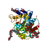


 PDBj
PDBj


 Assembly
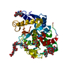

 Type: D-saccharide, alpha linking / Mass: 180.156 Da / Num. of mol.: 1


 Mass: 55.845 Da / Num. of mol.: 1 / Source method: obtained synthetically / Formula: Fe
 Mass: 60.009 Da / Num. of mol.: 1 / Source method: obtained synthetically / Formula: CO3
 Mass: 65.409 Da / Num. of mol.: 2 / Source method: obtained synthetically / Formula: Zn
 Mass: 96.063 Da / Num. of mol.: 1 / Source method: obtained synthetically / Formula: SO4
 Sample preparation
 Processing