 Movie
Movie Controller

[English] 日本語
 Yorodumi
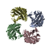
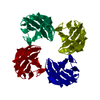
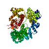
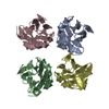
- PDB-2h4o: X-ray Crystal Structure of Protein yonK from Bacillus subtilis. N... -
+ Open data

- Basic information
| Entry | Database: PDB / ID: 2h4o | ||||||
|---|---|---|---|---|---|---|---|
| Title | X-ray Crystal Structure of Protein yonK from Bacillus subtilis. Northeast Structural Genomics Consortium Target SR415 | ||||||
 Components Components | YonK protein | ||||||
 Keywords Keywords | STRUCTURAL GENOMICS / UNKNOWN FUNCTION / PSI / PROTEIN STRUCTURE INITIATIVE / NORTHEAST STRUCTURAL GENOMICS CONSORTIUM / NESG / Bsu2107 (YonK protein) | ||||||
| Function / homology | OB fold (Dihydrolipoamide Acetyltransferase, E2P) - #10 / Bacillus phage SPbeta, YonK / YonK superfamily / YonK protein / OB fold (Dihydrolipoamide Acetyltransferase, E2P) / Other non-globular / Special / SPbeta prophage-derived uncharacterized protein YonK Function and homology information Function and homology information | ||||||
| Biological species |  | ||||||
| Method |  X-RAY DIFFRACTION / SYNCHROTRON / MAD / Resolution: 2.8 Å X-RAY DIFFRACTION / SYNCHROTRON / MAD / Resolution: 2.8 Å | ||||||
 Authors Authors | Seetharaman, J. / Sue, M. / Forouhar, F. / Ken, C. / Bonnie, C. / Ma, L. / Xiao, R. / Acton, T.B. / Hunt, J.F. / Tong, L. / Northeast Structural Genomics Consortium (NESG) | ||||||
 Citation Citation | Journal: To be Published Title: Crystal structure of the hypothetical protein from bacillus subtilis (yonk). Authors: Seetharaman, J. / Sue, M. / Forouhar, F. / Ken, C. / Bonnie, C. / Ma, L. / Xiao, R. / Acton, T.B. / Hunt, J.F. / Tong, L. / Northeast Structural Genomics Consortium (NESG) | ||||||
| History |
|
- Structure visualization
| Structure viewer | Molecule: MolmilJmol/JSmol |
|---|
- Downloads & links
-Download
| PDBx/mmCIF format | 2h4o.cif.gz | 62.6 KB | Display | PDBx/mmCIF format |
|---|---|---|---|---|
| PDB format | pdb2h4o.ent.gz | 47 KB | Display | PDB format |
| PDBx/mmJSON format | 2h4o.json.gz | Tree view | PDBx/mmJSON format | |
| Others |  Other downloads Other downloads |
-Validation report
| Arichive directory | https://data.pdbj.org/pub/pdb/validation_reports/h4/2h4oftp://data.pdbj.org/pub/pdb/validation_reports/h4/2h4o | HTTPS FTP |
|---|
-Related structure data
| Similar structure data | |
|---|---|
| Other databases |
-Links
 PDBj
PDBj- Assembly
| Deposited unit | 
| ||||||||
|---|---|---|---|---|---|---|---|---|---|
| 1 | 
| ||||||||
| Unit cell |
|
-Components
| #1: Protein | Mass: 9012.471 Da / Num. of mol.: 4 Source method: isolated from a genetically manipulated source Source: (gene. exp.) #2: Water | ChemComp-HOH / |  Mass: 18.015 Da / Num. of mol.: 28 / Source method: isolated from a natural source / Formula: H2O Mass: 18.015 Da / Num. of mol.: 28 / Source method: isolated from a natural source / Formula: H2OHas protein modification | Y | |
|---|
-Experimental details
-Experiment
| Experiment | Method: X-RAY DIFFRACTION / Number of used crystals: 1 |
|---|
- Sample preparation
| Crystal | Density Matthews: 2.26 Å3/Da / Density % sol: 45.68 % |
|---|---|
| Crystal grow | Temperature: 298 K / Method: vapor diffusion, hanging drop / pH: 9 Details: PEG 1000, 100mM TAPS pH 9.0, 120mM MgCl2, VAPOR DIFFUSION, HANGING DROP, temperature 298K |
-Data collection
| Diffraction | Mean temperature: 100 K | ||||||||||||
|---|---|---|---|---|---|---|---|---|---|---|---|---|---|
| Diffraction source | Source: SYNCHROTRON / Site: NSLS  / Beamline: X4A / Wavelength: 0.97913, 0.97941, 0.96780 / Beamline: X4A / Wavelength: 0.97913, 0.97941, 0.96780 | ||||||||||||
| Detector | Type: ADSC QUANTUM 4 / Detector: CCD / Date: Nov 24, 2005 / Details: Mirrors | ||||||||||||
| Radiation | Monochromator: SI 111 CHANNEL / Protocol: MAD / Monochromatic (M) / Laue (L): M / Scattering type: x-ray | ||||||||||||
| Radiation wavelength |
| ||||||||||||
| Reflection | Resolution: 2.8→50 Å / Num. all: 15558 / Num. obs: 15092 / % possible obs: 0.97 % / Observed criterion σ(F): 0 / Observed criterion σ(I): 0 / Redundancy: 4.2 % / Biso Wilson estimate: -0.3 Å2 / Rmerge(I) obs: 0.048 / Rsym value: 0.046 / Net I/σ(I): 17.7 | ||||||||||||
| Reflection shell | Resolution: 2.8→2.91 Å / Redundancy: 3.8 % / Rmerge(I) obs: 0.116 / Mean I/σ(I) obs: 16.1 / Num. unique all: 1704 / Rsym value: 0.181 / % possible all: 0.99 |
- Processing
| Software |
| |||||||||||||||||||||||||
|---|---|---|---|---|---|---|---|---|---|---|---|---|---|---|---|---|---|---|---|---|---|---|---|---|---|---|
| Refinement | Method to determine structure: MAD / Resolution: 2.8→28.34 Å / Rfactor Rfree error: 0.008 / Data cutoff high absF: 868587.01 / Data cutoff low absF: 0 / Isotropic thermal model: RESTRAINED / Cross valid method: THROUGHOUT / σ(F): 2 / Stereochemistry target values: Engh & Huber
| |||||||||||||||||||||||||
| Solvent computation | Solvent model: FLAT MODEL / Bsol: 44.8049 Å2 / ksol: 0.320902 e/Å3 | |||||||||||||||||||||||||
| Displacement parameters | Biso mean: 60.4 Å2
| |||||||||||||||||||||||||
| Refine analyze |
| |||||||||||||||||||||||||
| Refinement step | Cycle: LAST / Resolution: 2.8→28.34 Å
| |||||||||||||||||||||||||
| Refine LS restraints |
| |||||||||||||||||||||||||
| Refine LS restraints NCS | NCS model details: CONSTR | |||||||||||||||||||||||||
| LS refinement shell | Resolution: 2.8→2.98 Å / Rfactor Rfree error: 0.034 / Total num. of bins used: 6
| |||||||||||||||||||||||||
| Xplor file |
|
