 Movie
Movie Controller

[English] 日本語
 Yorodumi
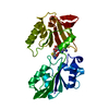
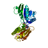
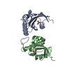
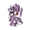
- PDB-2h2b: Crystal Structure of ZO-1 PDZ1 Bound to a Phage-Derived Ligand (W... -
+ Open data

- Basic information
| Entry | Database: PDB / ID: 2h2b | ||||||
|---|---|---|---|---|---|---|---|
| Title | Crystal Structure of ZO-1 PDZ1 Bound to a Phage-Derived Ligand (WRRTTYL) | ||||||
 Components Components | Tight junction protein ZO-1 | ||||||
 Keywords Keywords | CELL ADHESION / PDZ Domain / Phage Derived High Affinity Ligand | ||||||
| Function / homology |  Function and homology information Function and homology informationpositive regulation of blood-brain barrier permeability / adherens junction maintenance / ameloblast differentiation / RUNX1 regulates expression of components of tight junctions / positive regulation of cell-cell adhesion mediated by cadherin / establishment of endothelial intestinal barrier / SARS-CoV-2 targets PDZ proteins in cell-cell junction / protein localization to cell-cell junction / regulation of cell junction assembly / Regulation of gap junction activity ...positive regulation of blood-brain barrier permeability / adherens junction maintenance / ameloblast differentiation / RUNX1 regulates expression of components of tight junctions / positive regulation of cell-cell adhesion mediated by cadherin / establishment of endothelial intestinal barrier / SARS-CoV-2 targets PDZ proteins in cell-cell junction / protein localization to cell-cell junction / regulation of cell junction assembly / Regulation of gap junction activity / protein localization to bicellular tight junction / protein localization to adherens junction / gap junction / Apoptotic cleavage of cell adhesion proteins / negative regulation of stress fiber assembly / Signaling by Hippo / cell-cell junction organization / actomyosin structure organization / cell-cell junction assembly / podosome / tight junction / regulation of bicellular tight junction assembly / apical junction complex / positive regulation of sprouting angiogenesis / regulation of cytoskeleton organization / maintenance of blood-brain barrier / bicellular tight junction / cell adhesion molecule binding / adherens junction / cell-cell adhesion / apical part of cell / cell junction / actin cytoskeleton organization / basolateral plasma membrane / calmodulin binding / positive regulation of cell migration / cadherin binding / positive regulation of cell population proliferation / negative regulation of apoptotic process / protein-containing complex / plasma membrane / cytoplasm / cytosol Similarity search - Function | ||||||
| Biological species |  Homo sapiens (human) Homo sapiens (human) | ||||||
| Method |  X-RAY DIFFRACTION / SYNCHROTRON / MOLECULAR REPLACEMENT / Resolution: 1.6 Å X-RAY DIFFRACTION / SYNCHROTRON / MOLECULAR REPLACEMENT / Resolution: 1.6 Å | ||||||
 Authors Authors | Appleton, B.A. / Zhang, Y. / Wu, P. / Yin, J.P. / Hunziker, W. / Skelton, N.J. / Sidhu, S.S. / Wiesmann, C. | ||||||
 Citation Citation | Journal: J.Biol.Chem. / Year: 2006 Title: Comparative structural analysis of the Erbin PDZ domain and the first PDZ domain of ZO-1. Insights into determinants of PDZ domain specificity. Authors: Appleton, B.A. / Zhang, Y. / Wu, P. / Yin, J.P. / Hunziker, W. / Skelton, N.J. / Sidhu, S.S. / Wiesmann, C. #1: Journal: J.Biol.Chem. / Year: 2006 Title: Convergent and Divergent Ligand Specificity Amongst the PDZ Domains of the LAP and ZO Families Authors: Zhang, Y. / Yeh, S. / Appleton, B.A. / Held, H.A. / Kausalya, J.P. / Phua, D.C.Y. / Wong, W.L. / Lasky, L.A. / Wiesmann, C. / Hunziker, W. / Sidhu, S.S. | ||||||
| History |
|
- Structure visualization
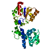
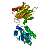
| Structure viewer | Molecule: MolmilJmol/JSmol |
|---|
- Downloads & links
-Download
| PDBx/mmCIF format | 2h2b.cif.gz | 37.6 KB | Display | PDBx/mmCIF format |
|---|---|---|---|---|
| PDB format | pdb2h2b.ent.gz | 24.7 KB | Display | PDB format |
| PDBx/mmJSON format | 2h2b.json.gz | Tree view | PDBx/mmJSON format | |
| Others |  Other downloads Other downloads |
-Validation report
| Arichive directory | https://data.pdbj.org/pub/pdb/validation_reports/h2/2h2bftp://data.pdbj.org/pub/pdb/validation_reports/h2/2h2b | HTTPS FTP |
|---|
-Related structure data
| Related structure data |  2h2cC  2h3lC 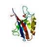 2h3mSC C: citing same article ( S: Starting model for refinement |
|---|---|
| Similar structure data |


-Links
 PDBj
PDBj


- Assembly
| Deposited unit | 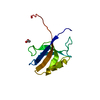
| ||||||||
|---|---|---|---|---|---|---|---|---|---|
| 1 | 
| ||||||||
| Unit cell |
| ||||||||
| Components on special symmetry positions |
|
-Components
| #1: Protein | Mass: 11716.234 Da / Num. of mol.: 1 / Fragment: First PDZ domain Source method: isolated from a genetically manipulated source Source: (gene. exp.) Homo sapiens (human) / Gene: ZO1 (TJP1) / Plasmid: pET22d / Species (production host): Escherichia coli / Production host:  |
|---|---|
| #2: Chemical | ChemComp-ACY /   Mass: 60.052 Da / Num. of mol.: 1 / Source method: obtained synthetically / Formula: C2H4O2 Mass: 60.052 Da / Num. of mol.: 1 / Source method: obtained synthetically / Formula: C2H4O2 |
| #3: Water | ChemComp-HOH /  Mass: 18.015 Da / Num. of mol.: 109 / Source method: isolated from a natural source / Formula: H2O Mass: 18.015 Da / Num. of mol.: 109 / Source method: isolated from a natural source / Formula: H2O |
-Experimental details
-Experiment
| Experiment | Method: X-RAY DIFFRACTION / Number of used crystals: 1 |
|---|
- Sample preparation
| Crystal | Density Matthews: 2.72 Å3/Da / Density % sol: 54.85 % |
|---|---|
| Crystal grow | Temperature: 292 K / Method: vapor diffusion, sitting drop / pH: 4.6 Details: 0.1 M Sodium Acetate, 0.2 M Ammonium Sulfate, 30% PEG 2000 Monomethyl Ether, pH 4.6, VAPOR DIFFUSION, SITTING DROP, temperature 292K |
-Data collection
| Diffraction | Mean temperature: 100 K | ||||||||||||||||||||||||||||||||||||||||||||||||||||||||||||||||||||||||||||||||||||||||
|---|---|---|---|---|---|---|---|---|---|---|---|---|---|---|---|---|---|---|---|---|---|---|---|---|---|---|---|---|---|---|---|---|---|---|---|---|---|---|---|---|---|---|---|---|---|---|---|---|---|---|---|---|---|---|---|---|---|---|---|---|---|---|---|---|---|---|---|---|---|---|---|---|---|---|---|---|---|---|---|---|---|---|---|---|---|---|---|---|---|
| Diffraction source | Source: SYNCHROTRON / Site: ALS  / Beamline: 5.0.1 / Wavelength: 1 Å / Beamline: 5.0.1 / Wavelength: 1 Å | ||||||||||||||||||||||||||||||||||||||||||||||||||||||||||||||||||||||||||||||||||||||||
| Detector | Type: ADSC QUANTUM 210 / Detector: CCD / Date: Sep 26, 2004 | ||||||||||||||||||||||||||||||||||||||||||||||||||||||||||||||||||||||||||||||||||||||||
| Radiation | Protocol: SINGLE WAVELENGTH / Monochromatic (M) / Laue (L): M / Scattering type: x-ray | ||||||||||||||||||||||||||||||||||||||||||||||||||||||||||||||||||||||||||||||||||||||||
| Radiation wavelength | Wavelength: 1 Å / Relative weight: 1 | ||||||||||||||||||||||||||||||||||||||||||||||||||||||||||||||||||||||||||||||||||||||||
| Reflection | Resolution: 1.61→50 Å / Num. obs: 17606 / % possible obs: 99.6 % / Redundancy: 7.3 % / Rsym value: 0.052 / Χ2: 1.024 / Net I/σ(I): 31.9 | ||||||||||||||||||||||||||||||||||||||||||||||||||||||||||||||||||||||||||||||||||||||||
| Reflection shell | Diffraction-ID: 1
|
- Processing
| Software |
| ||||||||||||||||||||||||||||||||||||||||||||||||||||||||||||||||||||||||||||||||||||||||||
|---|---|---|---|---|---|---|---|---|---|---|---|---|---|---|---|---|---|---|---|---|---|---|---|---|---|---|---|---|---|---|---|---|---|---|---|---|---|---|---|---|---|---|---|---|---|---|---|---|---|---|---|---|---|---|---|---|---|---|---|---|---|---|---|---|---|---|---|---|---|---|---|---|---|---|---|---|---|---|---|---|---|---|---|---|---|---|---|---|---|---|---|
| Refinement | Method to determine structure: MOLECULAR REPLACEMENT Starting model: PDB Entry: 2H3M Resolution: 1.6→30 Å / Cor.coef. Fo:Fc: 0.952 / Cor.coef. Fo:Fc free: 0.942 / SU B: 2.734 / SU ML: 0.051 / TLS residual ADP flag: LIKELY RESIDUAL / Cross valid method: THROUGHOUT / σ(F): 0 / ESU R: 0.09 / ESU R Free: 0.09 / Stereochemistry target values: MAXIMUM LIKELIHOOD / Details: HYDROGENS HAVE BEEN ADDED IN THE RIDING POSITIONS
| ||||||||||||||||||||||||||||||||||||||||||||||||||||||||||||||||||||||||||||||||||||||||||
| Solvent computation | Ion probe radii: 0.8 Å / Shrinkage radii: 0.8 Å / VDW probe radii: 1.2 Å / Solvent model: BABINET MODEL WITH MASK | ||||||||||||||||||||||||||||||||||||||||||||||||||||||||||||||||||||||||||||||||||||||||||
| Displacement parameters | Biso mean: 27.636 Å2
| ||||||||||||||||||||||||||||||||||||||||||||||||||||||||||||||||||||||||||||||||||||||||||
| Refinement step | Cycle: LAST / Resolution: 1.6→30 Å
| ||||||||||||||||||||||||||||||||||||||||||||||||||||||||||||||||||||||||||||||||||||||||||
| Refine LS restraints |
| ||||||||||||||||||||||||||||||||||||||||||||||||||||||||||||||||||||||||||||||||||||||||||
| LS refinement shell | Resolution: 1.604→1.646 Å / Total num. of bins used: 20
| ||||||||||||||||||||||||||||||||||||||||||||||||||||||||||||||||||||||||||||||||||||||||||
| Refinement TLS params. | Method: refined / Refine-ID: X-RAY DIFFRACTION
| ||||||||||||||||||||||||||||||||||||||||||||||||||||||||||||||||||||||||||||||||||||||||||
| Refinement TLS group | Refine-ID: X-RAY DIFFRACTION / Selection: ALL / Auth asym-ID: A / Label asym-ID: A
|
