 Movie
Movie Controller


+ Open data

- Basic information
| Entry | Database: PDB / ID: 2h0f | ||||||
|---|---|---|---|---|---|---|---|
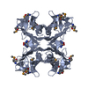
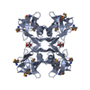
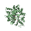
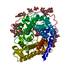
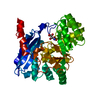
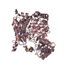
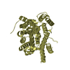
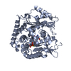
| Title | Crystal Structure of PucM in the presence of 8-azaxanthine | ||||||
 Components Components | Transthyretin-like protein pucM | ||||||
 Keywords Keywords | HYDROLASE / BETA SANDWITCH / INHIBITOR COMPLEX / HIU | ||||||
| Function / homology |  Function and homology information Function and homology informationhydroxyisourate hydrolase / hydroxyisourate hydrolase activity / urate catabolic process / purine nucleobase metabolic process / identical protein binding Similarity search - Function | ||||||
| Biological species |  | ||||||
| Method |  X-RAY DIFFRACTION / SYNCHROTRON / FOURIER SYNTHESIS / Resolution: 2.7 Å X-RAY DIFFRACTION / SYNCHROTRON / FOURIER SYNTHESIS / Resolution: 2.7 Å | ||||||
 Authors Authors | Rhee, S. | ||||||
 Citation Citation | Journal: Proc.Natl.Acad.Sci.Usa / Year: 2006 Title: Structural and functional analysis of PucM, a hydrolase in the ureide pathway and a member of the transthyretin-related protein family. Authors: Jung, D.-K. / Lee, Y. / Park, S.G. / Park, B.C. / Kim, G.-H. / Rhee, S. | ||||||
| History |
|
- Structure visualization
| Structure viewer | Molecule: MolmilJmol/JSmol |
|---|
- Downloads & links
-Download
| PDBx/mmCIF format | 2h0f.cif.gz | 59.3 KB | Display | PDBx/mmCIF format |
|---|---|---|---|---|
| PDB format | pdb2h0f.ent.gz | 43.5 KB | Display | PDB format |
| PDBx/mmJSON format | 2h0f.json.gz | Tree view | PDBx/mmJSON format | |
| Others |  Other downloads Other downloads |
-Validation report
| Arichive directory | https://data.pdbj.org/pub/pdb/validation_reports/h0/2h0fftp://data.pdbj.org/pub/pdb/validation_reports/h0/2h0f | HTTPS FTP |
|---|
-Related structure data


-Links
 PDBj
PDBj

- Assembly
| Deposited unit | 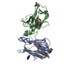
| |||||||||
|---|---|---|---|---|---|---|---|---|---|---|
| 1 | 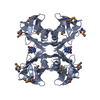
| |||||||||
| 2 | 
| |||||||||
| Unit cell |
| |||||||||
| Components on special symmetry positions |
| |||||||||
| Details | Chain A and B forms an indepedent crystallographic-symmetry-related homotetramer. |
-Components
| #1: Protein | Mass: 13703.716 Da / Num. of mol.: 2 Source method: isolated from a genetically manipulated source Source: (gene. exp.) #2: Chemical | ChemComp-AZA / |   Mass: 153.099 Da / Num. of mol.: 1 / Source method: obtained synthetically / Formula: C4H3N5O2 Mass: 153.099 Da / Num. of mol.: 1 / Source method: obtained synthetically / Formula: C4H3N5O2#3: Water | ChemComp-HOH / |  Mass: 18.015 Da / Num. of mol.: 84 / Source method: isolated from a natural source / Formula: H2O Mass: 18.015 Da / Num. of mol.: 84 / Source method: isolated from a natural source / Formula: H2OHas protein modification | Y | |
|---|
-Experimental details
-Experiment
| Experiment | Method: X-RAY DIFFRACTION / Number of used crystals: 1 |
|---|
- Sample preparation
| Crystal | Density Matthews: 3.52 Å3/Da / Density % sol: 65.1 % |
|---|---|
| Crystal grow | Temperature: 295 K / Method: vapor diffusion / pH: 4.2 Details: 0.1M sodium acetate, 0.5M NaCl, 0.5M ammonium sulfate, pH 4.2, VAPOR DIFFUSION, temperature 295K |
-Data collection
| Diffraction | Mean temperature: 100 K | ||||||||||||||||||||||||||||||||||||||||||||||||||||||||||||||||||
|---|---|---|---|---|---|---|---|---|---|---|---|---|---|---|---|---|---|---|---|---|---|---|---|---|---|---|---|---|---|---|---|---|---|---|---|---|---|---|---|---|---|---|---|---|---|---|---|---|---|---|---|---|---|---|---|---|---|---|---|---|---|---|---|---|---|---|---|
| Diffraction source | Source: SYNCHROTRON / Site: PAL/PLS  / Beamline: 6B / Wavelength: 0.97144 Å / Beamline: 6B / Wavelength: 0.97144 Å | ||||||||||||||||||||||||||||||||||||||||||||||||||||||||||||||||||
| Detector | Type: BRUKER PROTEUM 300 / Detector: CCD / Date: Oct 16, 2005 | ||||||||||||||||||||||||||||||||||||||||||||||||||||||||||||||||||
| Radiation | Protocol: SINGLE WAVELENGTH / Monochromatic (M) / Laue (L): M / Scattering type: x-ray | ||||||||||||||||||||||||||||||||||||||||||||||||||||||||||||||||||
| Radiation wavelength | Wavelength: 0.97144 Å / Relative weight: 1 | ||||||||||||||||||||||||||||||||||||||||||||||||||||||||||||||||||
| Reflection | Resolution: 2.6→30 Å / Num. all: 12724 / Num. obs: 12724 / % possible obs: 100 % / Observed criterion σ(F): 0 / Observed criterion σ(I): 0 / Redundancy: 13.5 % / Rmerge(I) obs: 0.131 / Χ2: 0.801 / Net I/σ(I): 6.3 | ||||||||||||||||||||||||||||||||||||||||||||||||||||||||||||||||||
| Reflection shell |
|
- Processing
| Software |
| |||||||||||||||||||||||||
|---|---|---|---|---|---|---|---|---|---|---|---|---|---|---|---|---|---|---|---|---|---|---|---|---|---|---|
| Refinement | Method to determine structure: FOURIER SYNTHESIS / Resolution: 2.7→30 Å / σ(F): 1109 / Stereochemistry target values: Engh & Huber
| |||||||||||||||||||||||||
| Solvent computation | Bsol: 26.267 Å2 | |||||||||||||||||||||||||
| Displacement parameters | Biso mean: 36.501 Å2
| |||||||||||||||||||||||||
| Refinement step | Cycle: LAST / Resolution: 2.7→30 Å
| |||||||||||||||||||||||||
| Refine LS restraints |
| |||||||||||||||||||||||||
| Xplor file |
|
