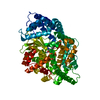
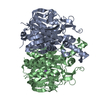
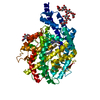
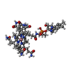
SIGNALING PROTEIN/MEMBRANE PROTEIN / OUTER-MEMBRANE ACTIVE TRANSPORT / BETA-BARREL / TONB / MEMBRANE PROTEIN / SIGNALING PROTEIN-MEMBRANE PROTEIN COMPLEX
Function / homology
Function and homology information
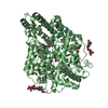
receptor-mediated bacteriophage irreversible attachment to host cell / colicin transport / ABC-type vitamin B12 transporter activity / energy transducer activity / Metal ion assimilation from the host / cobalamin transport / intracellular monoatomic cation homeostasis / siderophore transport / Iron assimilation using enterobactin / transmembrane transporter complex ...receptor-mediated bacteriophage irreversible attachment to host cell / colicin transport / ABC-type vitamin B12 transporter activity / energy transducer activity / Metal ion assimilation from the host / cobalamin transport / intracellular monoatomic cation homeostasis / siderophore transport / Iron assimilation using enterobactin / transmembrane transporter complex / plasma membrane protein complex / cell envelope / Heme assimilation / porin activity / pore complex / cell outer membrane / transmembrane transport / outer membrane-bounded periplasmic space / protein transport / monoatomic ion transmembrane transport / intracellular iron ion homeostasis / protein domain specific binding / calcium ion binding / membrane / plasma membrane Similarity search - Function
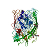
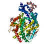
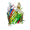
THE BIOLOGICAL ASSEMBLY IS A 1:1 COMPLEX OF BTUB AND TONB, WITH THE PHYSIOLOGICAL INTERFACE BETWEEN BTUB RESIDUES 6-12 AND TONB RESIDUES 158-171 AND 225-232.
In the structure databanks used in Yorodumi, some data are registered as the other names, "COVID-19 virus" and "2019-nCoV". Here are the details of the virus and the list of structure data.
Jan 31, 2019. EMDB accession codes are about to change! (news from PDBe EMDB page)
EMDB accession codes are about to change! (news from PDBe EMDB page)
The allocation of 4 digits for EMDB accession codes will soon come to an end. Whilst these codes will remain in use, new EMDB accession codes will include an additional digit and will expand incrementally as the available range of codes is exhausted. The current 4-digit format prefixed with “EMD-” (i.e. EMD-XXXX) will advance to a 5-digit format (i.e. EMD-XXXXX), and so on. It is currently estimated that the 4-digit codes will be depleted around Spring 2019, at which point the 5-digit format will come into force.
The EM Navigator/Yorodumi systems omit the EMD- prefix.
Related info.:Q: What is EMD? / ID/Accession-code notation in Yorodumi/EM Navigator
Yorodumi is a browser for structure data from EMDB, PDB, SASBDB, etc.
This page is also the successor to EM Navigator detail page, and also detail information page/front-end page for Omokage search.
The word "yorodu" (or yorozu) is an old Japanese word meaning "ten thousand". "mi" (miru) is to see.
Related info.:EMDB / PDB / SASBDB / Comparison of 3 databanks / Yorodumi Search / Aug 31, 2016. New EM Navigator & Yorodumi / Yorodumi Papers / Jmol/JSmol / Function and homology information / Changes in new EM Navigator and Yorodumi
 Movie
Movie Controller


 Open data
 Basic information
 Components
 Keywords
 Function and homology information
 X-RAY DIFFRACTION /
  Authors
 Citation
 Structure visualization
 Downloads & links
 Other downloads
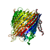
 PDBj
PDBj

 Assembly
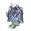


 Mass: 40.078 Da / Num. of mol.: 2 / Source method: obtained synthetically / Formula: Ca
 Mass: 1356.373 Da / Num. of mol.: 1 / Source method: obtained synthetically / Formula: C63H89CoN14O14P
 Mass: 229.402 Da / Num. of mol.: 4 / Source method: obtained synthetically / Formula: C14H31NO / Comment: LDAO, detergent*YM
 Mass: 114.229 Da / Num. of mol.: 3 / Source method: obtained synthetically / Formula: C8H18
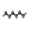
 Mass: 86.175 Da / Num. of mol.: 2 / Source method: obtained synthetically / Formula: C6H14
 Sample preparation
 / Beamline: 22-ID / Wavelength: 1
  Processing