 Movie
Movie Controller


+ Open data

- Basic information
| Entry | Database: PDB / ID: 2gpr | ||||||
|---|---|---|---|---|---|---|---|
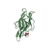
| Title | GLUCOSE PERMEASE IIA FROM MYCOPLASMA CAPRICOLUM | ||||||
 Components Components | GLUCOSE-PERMEASE IIA COMPONENT | ||||||
 Keywords Keywords | PHOSPHOTRANSFERASE / GLUCOSE PERMEASE / ENZYME IIA / MYCOPLASMA | ||||||
| Function / homology |  Function and homology information Function and homology informationphosphoenolpyruvate-dependent sugar phosphotransferase system / kinase activity / metal ion binding / cytoplasm Similarity search - Function | ||||||
| Biological species |  Mycoplasma capricolum (bacteria) Mycoplasma capricolum (bacteria) | ||||||
| Method |  X-RAY DIFFRACTION / MOLECULAR REPLACEMENT / Resolution: 2.5 Å X-RAY DIFFRACTION / MOLECULAR REPLACEMENT / Resolution: 2.5 Å | ||||||
 Authors Authors | Huang, K. / Herzberg, O. | ||||||
 Citation Citation | Journal: Structure / Year: 1998 Title: A promiscuous binding surface: crystal structure of the IIA domain of the glucose-specific permease from Mycoplasma capricolum. Authors: Huang, K. / Kapadia, G. / Zhu, P.P. / Peterkofsky, A. / Herzberg, O. | ||||||
| History |
|
- Structure visualization
| Structure viewer | Molecule: MolmilJmol/JSmol |
|---|
- Downloads & links
-Download
| PDBx/mmCIF format | 2gpr.cif.gz | 41.9 KB | Display | PDBx/mmCIF format |
|---|---|---|---|---|
| PDB format | pdb2gpr.ent.gz | 29.4 KB | Display | PDB format |
| PDBx/mmJSON format | 2gpr.json.gz | Tree view | PDBx/mmJSON format | |
| Others |  Other downloads Other downloads |
-Validation report
| Summary document | 2gpr_validation.pdf.gz | 366.7 KB | Display | wwPDB validaton report |
|---|---|---|---|---|
| Full document | 2gpr_full_validation.pdf.gz | 377.2 KB | Display | |
| Data in XML | 2gpr_validation.xml.gz | 6.3 KB | Display | |
| Data in CIF | 2gpr_validation.cif.gz | 8.8 KB | Display | |
| Arichive directory | https://data.pdbj.org/pub/pdb/validation_reports/gp/2gprftp://data.pdbj.org/pub/pdb/validation_reports/gp/2gpr | HTTPS FTP |
-Related structure data
| Related structure data |  1gprS S: Starting model for refinement |
|---|---|
| Similar structure data |


-Links
 PDBj
PDBj- Assembly
| Deposited unit | 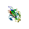
| ||||||||
|---|---|---|---|---|---|---|---|---|---|
| 1 |
| ||||||||
| Unit cell |
|
-Components
| #1: Protein | Mass: 16723.242 Da / Num. of mol.: 1 / Fragment: DOMAIN IIA Source method: isolated from a genetically manipulated source Source: (gene. exp.) Mycoplasma capricolum (bacteria) / Strain: KID / Cellular location: CYTOPLASM / Production host: References: UniProt: P45618, protein-Npi-phosphohistidine-sugar phosphotransferase |
|---|---|
| #2: Water | ChemComp-HOH /  Mass: 18.015 Da / Num. of mol.: 62 / Source method: isolated from a natural source / Formula: H2O Mass: 18.015 Da / Num. of mol.: 62 / Source method: isolated from a natural source / Formula: H2O |
-Experimental details
-Experiment
| Experiment | Method: X-RAY DIFFRACTION / Number of used crystals: 1 |
|---|
- Sample preparation
| Crystal | Density Matthews: 2.07 Å3/Da / Density % sol: 41 % | ||||||||||||||||||||||||||||||||||||
|---|---|---|---|---|---|---|---|---|---|---|---|---|---|---|---|---|---|---|---|---|---|---|---|---|---|---|---|---|---|---|---|---|---|---|---|---|---|
| Crystal grow | pH: 7.5 Details: PROTEIN WAS CRYSTALLIZED FROM 20-30% PEG 3000, 0.3MM ZNCL2, 100MM TRIS-HCL BUFFER, PH 7.5. | ||||||||||||||||||||||||||||||||||||
| Crystal | *PLUS | ||||||||||||||||||||||||||||||||||||
| Crystal grow | *PLUS Method: vapor diffusion, hanging drop | ||||||||||||||||||||||||||||||||||||
| Components of the solutions | *PLUS
|
-Data collection
| Diffraction | Mean temperature: 298 K |
|---|---|
| Diffraction source | Source: ROTATING ANODE / Type: RIGAKU RUH2R / Wavelength: 1.5418 |
| Detector | Type: SIEMENS / Detector: AREA DETECTOR / Date: Nov 1, 1995 |
| Radiation | Monochromator: NI FILTER / Monochromatic (M) / Laue (L): M / Scattering type: x-ray |
| Radiation wavelength | Wavelength: 1.5418 Å / Relative weight: 1 |
| Reflection | Resolution: 2.5→20 Å / Num. obs: 4952 / % possible obs: 97 % / Observed criterion σ(I): 2 / Redundancy: 2.2 % / Rsym value: 0.069 / Net I/σ(I): 28.3 |
| Reflection | *PLUS Num. measured all: 12034 / Rmerge(I) obs: 0.063 |
| Reflection shell | *PLUS Highest resolution: 2.5 Å / Lowest resolution: 2.66 Å / % possible obs: 85.3 % / Rmerge(I) obs: 0.162 / Mean I/σ(I) obs: 6.1 |
- Processing
| Software |
| ||||||||||||||||||||||||||||||
|---|---|---|---|---|---|---|---|---|---|---|---|---|---|---|---|---|---|---|---|---|---|---|---|---|---|---|---|---|---|---|---|
| Refinement | Method to determine structure: MOLECULAR REPLACEMENT Starting model: PDB ENTRY 1GPR Resolution: 2.5→20 Å / Isotropic thermal model: TNT BCORREL / σ(F): 2 / Stereochemistry target values: TNT PROTGEO /
| ||||||||||||||||||||||||||||||
| Solvent computation | Solvent model: BABINET SCALING / Bsol: 134.2 Å2 / ksol: 0.6 e/Å3 | ||||||||||||||||||||||||||||||
| Refinement step | Cycle: LAST / Resolution: 2.5→20 Å
| ||||||||||||||||||||||||||||||
| Refine LS restraints |
| ||||||||||||||||||||||||||||||
| Software | *PLUS Name: TNT / Version: V5.0 / Classification: refinement | ||||||||||||||||||||||||||||||
| Refine LS restraints | *PLUS
|
