 Movie
Movie Controller

[English] 日本語
 Yorodumi
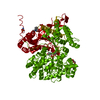
- PDB-2gpa: ALLOSTERIC INHIBITION OF GLYCOGEN PHOSPHORYLASE A BY A POTENTIAL ... -
+ Open data

- Basic information
| Entry | Database: PDB / ID: 2gpa | ||||||
|---|---|---|---|---|---|---|---|
| Title | ALLOSTERIC INHIBITION OF GLYCOGEN PHOSPHORYLASE A BY A POTENTIAL ANTIDIABETIC DRUG | ||||||
 Components Components | PROTEIN (GLYCOGEN PHOSPHORYLASE) | ||||||
 Keywords Keywords | TRANSFERASE / DIABETES / GLYCOGEN METABOLISM / PHOSPHORYLASE A / INHIBITION / ALLOSTERIC SITE | ||||||
| Function / homology |  Function and homology information Function and homology informationglycogen phosphorylase / glycogen phosphorylase activity / glycogen catabolic process / skeletal muscle myofibril / pyridoxal phosphate binding / nucleotide binding Similarity search - Function | ||||||
| Biological species |  | ||||||
| Method |  X-RAY DIFFRACTION / SYNCHROTRON / OTHER / Resolution: 2 Å X-RAY DIFFRACTION / SYNCHROTRON / OTHER / Resolution: 2 Å | ||||||
 Authors Authors | Oikonomakos, N.G. / Tsitsanou, K.E. / Zographos, S.E. / Skamnaki, V.T. | ||||||
 Citation Citation | Journal: Protein Sci. / Year: 1999 Title: Allosteric inhibition of glycogen phosphorylase a by the potential antidiabetic drug 3-isopropyl 4-(2-chlorophenyl)-1,4-dihydro-1-ethyl-2-methyl-pyridine-3,5,6-tricarbo xylate. Authors: Oikonomakos, N.G. / Tsitsanou, K.E. / Zographos, S.E. / Skamnaki, V.T. / Goldmann, S. / Bischoff, H. #1: Journal: To be PublishedTitle: Effects of Commonly Used Cryoprotectants on Glycogen Phosphorylase Activity and Structure Authors: Tsitsanou, K.E. / Oikonomakos, N.G. / Zographos, S.E. / Skamnaki, V.T. / Gregoriou, M. / Watson, K.A. / Johnson, L.N. / Fleet, G.W.J. #2: Journal: Structure / Year: 1997Title: The Structure of Glycogen Phosphorylase B with an Alkyl-Dihydropyridine- Dicarboxylic Acid Compound, a Novel and Potent Inhibitor Authors: Zographos, S.E. / Oikonomakos, N.G. / Tsitsanou, K.E. / Leonidas, D.D. / Chrysina, E.D. / Skamnaki, V.T. / Bischoff, H. / Goldman, S. / Schramm, M. / Watson, K.A. / Johnson, L.N. | ||||||
| History |
|
- Structure visualization
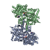
| Structure viewer | Molecule: MolmilJmol/JSmol |
|---|
- Downloads & links
-Download
| PDBx/mmCIF format | 2gpa.cif.gz | 192.6 KB | Display | PDBx/mmCIF format |
|---|---|---|---|---|
| PDB format | pdb2gpa.ent.gz | 152 KB | Display | PDB format |
| PDBx/mmJSON format | 2gpa.json.gz | Tree view | PDBx/mmJSON format | |
| Others |  Other downloads Other downloads |
-Validation report
| Arichive directory | https://data.pdbj.org/pub/pdb/validation_reports/gp/2gpaftp://data.pdbj.org/pub/pdb/validation_reports/gp/2gpa | HTTPS FTP |
|---|
-Related structure data


-Links
 PDBj
PDBj


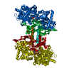
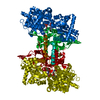
- Assembly
| Deposited unit | 
| ||||||||
|---|---|---|---|---|---|---|---|---|---|
| 1 | 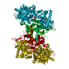
| ||||||||
| Unit cell |
|
-Components
| #1: Protein | Mass: 97371.180 Da / Num. of mol.: 1 / Source method: isolated from a natural source / Source: (natural) |
|---|---|
| #2: Sugar | ChemComp-GLC /   Type: D-saccharide, alpha linking / Mass: 180.156 Da / Num. of mol.: 1 Type: D-saccharide, alpha linking / Mass: 180.156 Da / Num. of mol.: 1Source method: isolated from a genetically manipulated source Formula: C6H12O6 |
| #3: Chemical | ChemComp-PLP /   Mass: 247.142 Da / Num. of mol.: 1 / Source method: obtained synthetically / Formula: C8H10NO6P Mass: 247.142 Da / Num. of mol.: 1 / Source method: obtained synthetically / Formula: C8H10NO6P |
| #4: Chemical | ChemComp-GOL /   Mass: 92.094 Da / Num. of mol.: 1 / Source method: obtained synthetically / Formula: C3H8O3 Mass: 92.094 Da / Num. of mol.: 1 / Source method: obtained synthetically / Formula: C3H8O3 |
| #5: Water | ChemComp-HOH /  Mass: 18.015 Da / Num. of mol.: 807 / Source method: isolated from a natural source / Formula: H2O Mass: 18.015 Da / Num. of mol.: 807 / Source method: isolated from a natural source / Formula: H2O |
-Experimental details
-Experiment
| Experiment | Method: X-RAY DIFFRACTION / Number of used crystals: 1 |
|---|
- Sample preparation
| Crystal | Density Matthews: 2.41 Å3/Da / Density % sol: 48 % |
|---|---|
| Crystal grow | pH: 6.7 / Details: pH 6.7 |
| Crystal grow | *PLUS Method: other / Details: Fletterick, R.J., (1976) J. Mol. Biol., 103, 1. |
-Data collection
| Diffraction | Mean temperature: 100 K |
|---|---|
| Diffraction source | Source: SYNCHROTRON / Site: ELETTRA  / Beamline: 5.2R / Wavelength: 1 / Beamline: 5.2R / Wavelength: 1 |
| Detector | Type: MAR scanner 180 mm plate / Detector: IMAGE PLATE / Date: Jul 1, 1997 |
| Radiation | Protocol: SINGLE WAVELENGTH / Monochromatic (M) / Laue (L): M / Scattering type: x-ray |
| Radiation wavelength | Wavelength: 1 Å / Relative weight: 1 |
| Reflection | Resolution: 2→13 Å / Num. obs: 57879 / % possible obs: 90.1 % / Redundancy: 7.5 % / Rmerge(I) obs: 0.079 / Net I/σ(I): 14.6 |
| Reflection shell | Resolution: 2→2.03 Å / Rmerge(I) obs: 0.41 / Mean I/σ(I) obs: 5 / % possible all: 92.6 |
| Reflection | *PLUS Lowest resolution: 13 Å / Num. measured all: 448708 |
| Reflection shell | *PLUS % possible obs: 92.6 % |
- Processing
| Software |
| ||||||||||||||||||||||||||||||||||||||||||||||||||||||||||||
|---|---|---|---|---|---|---|---|---|---|---|---|---|---|---|---|---|---|---|---|---|---|---|---|---|---|---|---|---|---|---|---|---|---|---|---|---|---|---|---|---|---|---|---|---|---|---|---|---|---|---|---|---|---|---|---|---|---|---|---|---|---|
| Refinement | Method to determine structure: OTHER / Resolution: 2→10 Å / Cross valid method: R FREE / σ(F): 0
| ||||||||||||||||||||||||||||||||||||||||||||||||||||||||||||
| Refinement step | Cycle: LAST / Resolution: 2→10 Å
| ||||||||||||||||||||||||||||||||||||||||||||||||||||||||||||
| Refine LS restraints |
| ||||||||||||||||||||||||||||||||||||||||||||||||||||||||||||
| Software | *PLUS Name: X-PLOR / Version: 3.8 / Classification: refinement | ||||||||||||||||||||||||||||||||||||||||||||||||||||||||||||
| Refinement | *PLUS Highest resolution: 2 Å / Lowest resolution: 10 Å / σ(F): 0 / Num. reflection Rfree: 2947 / % reflection Rfree: 5 % / Rfactor obs: 0.179 / Rfactor Rfree: 0.23 | ||||||||||||||||||||||||||||||||||||||||||||||||||||||||||||
| Solvent computation | *PLUS | ||||||||||||||||||||||||||||||||||||||||||||||||||||||||||||
| Displacement parameters | *PLUS | ||||||||||||||||||||||||||||||||||||||||||||||||||||||||||||
| Refine LS restraints | *PLUS
|
