 Movie
Movie Controller

[English] 日本語
 Yorodumi
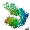
- PDB-2go5: Structure of signal recognition particle receptor (SR) in complex... -
+ Open data

- Basic information
| Entry | Database: PDB / ID: 2go5 | ||||||
|---|---|---|---|---|---|---|---|
| Title | Structure of signal recognition particle receptor (SR) in complex with signal recognition particle (SRP) and ribosome nascent chain complex | ||||||
 Components Components |
| ||||||
 Keywords Keywords | TRANSLATION/RNA / SR / SRP / RIBOSOME / TRANSLATION-RNA COMPLEX | ||||||
| Function / homology |  Function and homology information Function and homology informationSRP-dependent cotranslational protein targeting to membrane / signal recognition particle receptor complex / SRP-dependent cotranslational protein targeting to membrane, signal sequence recognition / endoplasmic reticulum signal peptide binding / signal recognition particle, endoplasmic reticulum targeting / granulocyte differentiation / signal recognition particle binding / cotranslational protein targeting to membrane / protein targeting to ER / signal-recognition-particle GTPase ...SRP-dependent cotranslational protein targeting to membrane / signal recognition particle receptor complex / SRP-dependent cotranslational protein targeting to membrane, signal sequence recognition / endoplasmic reticulum signal peptide binding / signal recognition particle, endoplasmic reticulum targeting / granulocyte differentiation / signal recognition particle binding / cotranslational protein targeting to membrane / protein targeting to ER / signal-recognition-particle GTPase / SRP-dependent cotranslational protein targeting to membrane, translocation / XBP1(S) activates chaperone genes / 7S RNA binding / exocrine pancreas development / SRP-dependent cotranslational protein targeting to membrane / ribonucleoprotein complex binding / cytoplasmic microtubule / neutrophil chemotaxis / maturation of LSU-rRNA from tricistronic rRNA transcript (SSU-rRNA, 5.8S rRNA, LSU-rRNA) / chloroplast / intracellular protein transport / GDP binding / cytosolic large ribosomal subunit / rRNA binding / nuclear speck / ribosome / structural constituent of ribosome / translation / ribonucleoprotein complex / GTPase activity / mRNA binding / endoplasmic reticulum membrane / GTP binding / nucleolus / endoplasmic reticulum / ATP hydrolysis activity / RNA binding / extracellular exosome / nucleoplasm / nucleus / membrane / cytosol Similarity search - Function | ||||||
| Biological species |   Homo sapiens (human) Homo sapiens (human) | ||||||
| Method | ELECTRON MICROSCOPY / single particle reconstruction / cryo EM / Resolution: 7.4 Å | ||||||
 Authors Authors | Halic, M. / Gartmann, M. / Schlenker, O. / Mielke, T. / Pool, M.R. / Sinning, I. / Beckmann, R. | ||||||
 Citation Citation | Journal: Science / Year: 2006 Title: Signal recognition particle receptor exposes the ribosomal translocon binding site. Authors: Mario Halic / Marco Gartmann / Oliver Schlenker / Thorsten Mielke / Martin R Pool / Irmgard Sinning / Roland Beckmann /  Abstract: Signal sequences of secretory and membrane proteins are recognized by the signal recognition particle (SRP) as they emerge from the ribosome. This results in their targeting to the membrane by ...Signal sequences of secretory and membrane proteins are recognized by the signal recognition particle (SRP) as they emerge from the ribosome. This results in their targeting to the membrane by docking with the SRP receptor, which facilitates transfer of the ribosome to the translocon. Here, we present the 8 angstrom cryo-electron microscopy structure of a "docking complex" consisting of a SRP-bound 80S ribosome and the SRP receptor. Interaction of the SRP receptor with both SRP and the ribosome rearranged the S domain of SRP such that a ribosomal binding site for the translocon, the L23e/L35 site, became exposed, whereas Alu domain-mediated elongation arrest persisted. | ||||||
| History |
| ||||||
| Remark 999 | SEQUENCE Residue (A G 124) and Residue (A G 125) are not linked. Distance of O3*-P bond is 4.22. ...SEQUENCE Residue (A G 124) and Residue (A G 125) are not linked. Distance of O3*-P bond is 4.22. Residue (A C 221) and Residue (A G 222) are not linked. Distance of O3*-P bond is 3.26. Residue (1 SER 0) and Residue (1 MET 1) are not linked. Distance of C-N bond is 17.58. Residue (1 GLY 27) and Residue (1 PRO 28) are not linked. Distance of C-N bond is 7.21. Residue (1 LEU 52) and Residue (1 THR 53) are not linked. Distance of C-N bond is 5.58. |
- Structure visualization
| Movie |
Movie viewer |
|---|---|
| Structure viewer | Molecule: MolmilJmol/JSmol |
- Downloads & links
-Download
| PDBx/mmCIF format | 2go5.cif.gz | 288.5 KB | Display | PDBx/mmCIF format |
|---|---|---|---|---|
| PDB format | pdb2go5.ent.gz | 215.2 KB | Display | PDB format |
| PDBx/mmJSON format | 2go5.json.gz | Tree view | PDBx/mmJSON format | |
| Others |  Other downloads Other downloads |
-Validation report
| Summary document | 2go5_validation.pdf.gz | 910.6 KB | Display | wwPDB validaton report |
|---|---|---|---|---|
| Full document | 2go5_full_validation.pdf.gz | 981.6 KB | Display | |
| Data in XML | 2go5_validation.xml.gz | 41.4 KB | Display | |
| Data in CIF | 2go5_validation.cif.gz | 61.7 KB | Display | |
| Arichive directory | https://data.pdbj.org/pub/pdb/validation_reports/go/2go5ftp://data.pdbj.org/pub/pdb/validation_reports/go/2go5 | HTTPS FTP |
-Related structure data
| Related structure data | 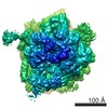 1217MC M: map data used to model this data C: citing same article ( |
|---|---|
| Similar structure data |
-Links
 PDBj
PDBj


- Assembly
| Deposited unit | 
|
|---|---|
| 1 |
|
-Components
-RNA chain , 2 types, 2 molecules A9
| #1: RNA chain | Mass: 41199.570 Da / Num. of mol.: 1 / Source method: isolated from a natural source / Source: (natural) |
|---|---|
| #2: RNA chain | Mass: 29170.479 Da / Num. of mol.: 1 / Source method: isolated from a natural source / Source: (natural) |
-Signal recognition particle ... , 4 types, 4 molecules BW12
| #3: Protein | Mass: 12561.688 Da / Num. of mol.: 1 Source method: isolated from a genetically manipulated source Source: (gene. exp.) |
|---|---|
| #4: Protein | Mass: 12473.440 Da / Num. of mol.: 1 Source method: isolated from a genetically manipulated source Source: (gene. exp.) |
| #5: Protein | Mass: 21329.512 Da / Num. of mol.: 1 Source method: isolated from a genetically manipulated source Source: (gene. exp.) Homo sapiens (human) / References: UniProt: P08240 |
| #6: Protein | Mass: 23536.982 Da / Num. of mol.: 1 Source method: isolated from a genetically manipulated source Source: (gene. exp.) |
-Ribosomal protein ... , 3 types, 3 molecules 546
| #7: Protein | Mass: 14401.041 Da / Num. of mol.: 1 Source method: isolated from a genetically manipulated source Source: (gene. exp.) |
|---|---|
| #8: Protein | Mass: 16920.203 Da / Num. of mol.: 1 Source method: isolated from a genetically manipulated source Source: (gene. exp.) |
| #9: Protein | Mass: 14152.365 Da / Num. of mol.: 1 Source method: isolated from a genetically manipulated source Source: (gene. exp.) |
-Details
| Has protein modification | Y |
|---|
-Experimental details
-Experiment
| Experiment | Method: ELECTRON MICROSCOPY |
|---|---|
| EM experiment | Aggregation state: PARTICLE / 3D reconstruction method: single particle reconstruction |
- Sample preparation
| Component | Name: SR-SRP-RNC COMPLEX / Type: RIBOSOME |
|---|---|
| Specimen | Embedding applied: NO / Shadowing applied: NO / Staining applied: NO / Vitrification applied: YES |
| Specimen support | Details: CARBON |
| Vitrification | Cryogen name: ETHANE |
- Electron microscopy imaging
| Experimental equipment |  Model: Tecnai F30 / Image courtesy: FEI Company |
|---|---|
| Microscopy | Model: FEI TECNAI F30 |
| Electron gun | Electron source:  FIELD EMISSION GUN / Accelerating voltage: 300 kV / Illumination mode: FLOOD BEAM FIELD EMISSION GUN / Accelerating voltage: 300 kV / Illumination mode: FLOOD BEAM |
| Electron lens | Mode: BRIGHT FIELD / Nominal defocus max: 3500 nm / Nominal defocus min: 800 nm |
| Image recording | Film or detector model: KODAK SO-163 FILM |
| Radiation | Protocol: SINGLE WAVELENGTH / Monochromatic (M) / Laue (L): M |
| Radiation wavelength | Relative weight: 1 |
- Processing
| Symmetry | Point symmetry: C1 (asymmetric) | ||||||||||||
|---|---|---|---|---|---|---|---|---|---|---|---|---|---|
| 3D reconstruction | Resolution: 7.4 Å / Resolution method: FSC 0.5 CUT-OFF / Symmetry type: POINT | ||||||||||||
| Refinement | Highest resolution: 7.4 Å | ||||||||||||
| Refinement step | Cycle: LAST / Highest resolution: 7.4 Å
|
