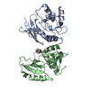
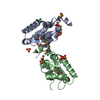
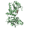
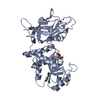
SEQUENCE: THE CONSTRUCT WAS EXPRESSED WITH A PURIFICATION TAG MGSDKIHHHHHHENLYFQG. THE TAG WAS ...SEQUENCE: THE CONSTRUCT WAS EXPRESSED WITH A PURIFICATION TAG MGSDKIHHHHHHENLYFQG. THE TAG WAS REMOVED WITH TEV PROTEASE LEAVING ONLY A GLYCINE (0) FOLLOWED BY RESIDUES 9-312 OF THE TM0771 SEQUENCE. A SMALL AMOUNT OF PROTEIN WITH THE TAG UNCLEAVED REMAINED IN THE SAMPLE AFTER PURIFICATION. A SERIES OF DIFFERENT LENGTH N-TERMINAL TRUNCATIONS WERE TRIED TO IMPROVE DIFFRACTION QUALITY FROM THIS TARGET. THE CONSTRUCT WITH RESIDUES 1-8 ELIMINATED PRODUCED THE BEST DIFFRACTING CRYSTAL.
Monochromator: Single crystal Si(111) bent monochromator (horizontal focusing) Protocol: MAD / Monochromatic (M) / Laue (L): M / Scattering type: x-ray
Radiation wavelength
ID
Wavelength (Å)
Relative weight
1
0.979224
1
2
0.91837
1
3
0.97894
1
Reflection
Resolution: 2→27.9 Å / Num. obs: 22105 / % possible obs: 94.7 % / Biso Wilson estimate: 42.314 Å2 / Rmerge(I) obs: 0.041 / Net I/σ(I): 11.88
Reflection shell
Diffraction-ID: 1
Resolution (Å)
Highest resolution (Å)
Rmerge(I) obs
Mean I/σ(I) obs
Num. measured obs
Num. unique obs
% possible all
2-2.07
0.415
2
6860
3580
83.8
2.07-2.15
0.314
2.7
7034
3711
90.1
2.15-2.25
0.251
3.3
7757
4060
92.2
2.25-2.37
0.184
4.5
7788
4072
93.6
2.37-2.52
0.125
6.3
7968
4166
97
2.52-2.71
0.093
8.3
7792
4088
97.8
2.71-2.99
0.067
10.9
8351
4353
98.7
2.99-3.42
0.046
15.5
8021
4231
99.7
3.42
0.025
25.1
7896
4199
99.1
-
Phasing
Phasing
Method: MAD
-
Processing
Software
Name
Version
Classification
NB
REFMAC
5.2.0005
refinement
XSCALE
datascaling
PDB_EXTRACT
1.701
dataextraction
XDS
datareduction
SHARP
V. 1
phasing
Refinement
Method to determine structure: MAD / Resolution: 2→27.94 Å / Cor.coef. Fo:Fc: 0.943 / Cor.coef. Fo:Fc free: 0.907 / SU B: 10.889 / SU ML: 0.153 / TLS residual ADP flag: LIKELY RESIDUAL / Cross valid method: THROUGHOUT / σ(F): 0 / ESU R: 0.217 / ESU R Free: 0.197 Stereochemistry target values: MAXIMUM LIKELIHOOD WITH PHASES Details: 1). HYDROGENS HAVE BEEN ADDED IN THE RIDING POSITIONS. 2). A MET-INHIBITION PROTOCOL WAS USED FOR SELENOMETHIONINE INCORPORATION DURING PROTEIN EXPRESSION. THE OCCUPANCY OF THE SE ATOMS IN ...Details: 1). HYDROGENS HAVE BEEN ADDED IN THE RIDING POSITIONS. 2). A MET-INHIBITION PROTOCOL WAS USED FOR SELENOMETHIONINE INCORPORATION DURING PROTEIN EXPRESSION. THE OCCUPANCY OF THE SE ATOMS IN THE MSE RESIDUES WAS REDUCED TO 0.75 TO ACCOUNT FOR THE REDUCED SCATTERING POWER DUE TO PARTIAL S-MET INCORPORATION. 3). TWO MOLECULES OF 2-ETHOXYETHANOL FROM THE CRYSTALLIZATION BUFFER HAS BEEN MODELED INTO THE STRUCTURE. 4). AN UNKNOWN LIGAND HAS BEEN MODELED INTO THE STRUCTURE BETWEEN THE SIDECHAINS OF LEU 168 AND ARG 254. 5.) ATOM RECORD CONTAINS RESIDUAL B FACTORS ONLY.
Rfactor
Num. reflection
% reflection
Selection details
Rfree
0.278
1130
5.1 %
RANDOM
Rwork
0.22
-
-
-
obs
0.223
22103
99.26 %
-
Solvent computation
Ion probe radii: 0.8 Å / Shrinkage radii: 0.8 Å / VDW probe radii: 1.2 Å / Solvent model: BABINET MODEL WITH MASK
Displacement parameters
Biso mean: 34.573 Å2
Baniso -1
Baniso -2
Baniso -3
1-
-0.96 Å2
0 Å2
0 Å2
2-
-
-0.96 Å2
0 Å2
3-
-
-
1.91 Å2
Refinement step
Cycle: LAST / Resolution: 2→27.94 Å
Protein
Nucleic acid
Ligand
Solvent
Total
Num. atoms
2346
0
17
107
2470
Refine LS restraints
Refine-ID
Type
Dev ideal
Dev ideal target
Number
X-RAY DIFFRACTION
r_bond_refined_d
0.013
0.022
2466
X-RAY DIFFRACTION
r_bond_other_d
0.003
0.02
2343
X-RAY DIFFRACTION
r_angle_refined_deg
1.684
1.989
3341
X-RAY DIFFRACTION
r_angle_other_deg
1.188
3
5432
X-RAY DIFFRACTION
r_dihedral_angle_1_deg
2.099
5
309
X-RAY DIFFRACTION
r_dihedral_angle_2_deg
25.182
24.112
107
X-RAY DIFFRACTION
r_dihedral_angle_3_deg
8.319
15
458
X-RAY DIFFRACTION
r_dihedral_angle_4_deg
8.252
15
18
X-RAY DIFFRACTION
r_chiral_restr
0.107
0.2
387
X-RAY DIFFRACTION
r_gen_planes_refined
0.006
0.02
2707
X-RAY DIFFRACTION
r_gen_planes_other
0.002
0.02
487
X-RAY DIFFRACTION
r_nbd_refined
0.18
0.3
532
X-RAY DIFFRACTION
r_nbd_other
0.123
0.3
2275
X-RAY DIFFRACTION
r_nbtor_refined
0.165
0.5
1206
X-RAY DIFFRACTION
r_nbtor_other
0.081
0.5
1307
X-RAY DIFFRACTION
r_xyhbond_nbd_refined
0.169
0.5
174
X-RAY DIFFRACTION
r_symmetry_vdw_refined
0.145
0.3
11
X-RAY DIFFRACTION
r_symmetry_vdw_other
0.109
0.3
55
X-RAY DIFFRACTION
r_symmetry_hbond_refined
0.22
0.5
17
X-RAY DIFFRACTION
r_mcbond_it
1.427
3
1513
X-RAY DIFFRACTION
r_mcbond_other
0.385
3
605
X-RAY DIFFRACTION
r_mcangle_it
2.42
5
2458
X-RAY DIFFRACTION
r_scbond_it
4.208
8
977
X-RAY DIFFRACTION
r_scangle_it
6.14
11
881
LS refinement shell
Resolution: 2.001→2.053 Å / Total num. of bins used: 20
Rfactor
Num. reflection
% reflection
Rfree
0.306
95
-
Rwork
0.257
1504
-
obs
-
1599
99.13 %
Refinement TLS params.
Method: refined / Refine-ID: X-RAY DIFFRACTION
ID
L11 (°2)
L12 (°2)
L13 (°2)
L22 (°2)
L23 (°2)
L33 (°2)
S11 (Å °)
S12 (Å °)
S13 (Å °)
S21 (Å °)
S22 (Å °)
S23 (Å °)
S31 (Å °)
S32 (Å °)
S33 (Å °)
T11 (Å2)
T12 (Å2)
T13 (Å2)
T22 (Å2)
T23 (Å2)
T33 (Å2)
Origin x (Å)
Origin y (Å)
Origin z (Å)
1
2.2739
3.3447
-0.0458
6.0604
-2.508
6.0805
-0.4766
0.8218
0.132
-0.865
0.6166
-0.1455
0.0443
-1.0041
-0.1401
-0.021
-0.0383
-0.0224
0.1935
0.0757
-0.0947
19.296
78.5452
27.1236
2
2.9919
1.5443
-2.4187
4.5978
-2.2967
4.9265
-0.093
-0.0827
0.0972
0.1565
0.0308
0.1339
0.2259
-0.3391
0.0622
0.0123
-0.0268
0.0039
0.0088
-0.0152
-0.0353
22.375
72.9649
39.6628
3
3.7286
-1.5147
0.092
1.6663
0.2696
0.7421
0.2961
0.1797
0.1402
-0.444
-0.2409
-0.1179
0.3113
-0.1436
-0.0552
0.2185
-0.0196
0.1473
-0.0902
-0.0095
-0.0867
36.5658
63.0713
26.8708
4
2.4266
0.6897
-0.4104
3.9158
0.159
1.8175
0.1276
-0.0452
0.0343
0.0263
0.0868
0.1446
-0.0776
-0.0887
-0.2144
0.0163
0.0281
0.0464
-0.0564
-0.0088
-0.0097
44.1169
44.9327
36.3526
Refinement TLS group
Refine-ID: X-RAY DIFFRACTION / Selection: ALL / Auth asym-ID: A / Label asym-ID: A
ID
Refine TLS-ID
Auth seq-ID
Label seq-ID
1
1
11 - 29
4 - 22
2
2
30 - 125
23 - 118
3
3
126 - 197
119 - 190
4
4
198 - 306
191 - 299
+
About Yorodumi
-
News
-
Feb 9, 2022. New format data for meta-information of EMDB entries
New format data for meta-information of EMDB entries
Version 3 of the EMDB header file is now the official format.
The previous official version 1.9 will be removed from the archive.
In the structure databanks used in Yorodumi, some data are registered as the other names, "COVID-19 virus" and "2019-nCoV". Here are the details of the virus and the list of structure data.
Jan 31, 2019. EMDB accession codes are about to change! (news from PDBe EMDB page)
EMDB accession codes are about to change! (news from PDBe EMDB page)
The allocation of 4 digits for EMDB accession codes will soon come to an end. Whilst these codes will remain in use, new EMDB accession codes will include an additional digit and will expand incrementally as the available range of codes is exhausted. The current 4-digit format prefixed with “EMD-” (i.e. EMD-XXXX) will advance to a 5-digit format (i.e. EMD-XXXXX), and so on. It is currently estimated that the 4-digit codes will be depleted around Spring 2019, at which point the 5-digit format will come into force.
The EM Navigator/Yorodumi systems omit the EMD- prefix.
Related info.:Q: What is EMD? / ID/Accession-code notation in Yorodumi/EM Navigator
Yorodumi is a browser for structure data from EMDB, PDB, SASBDB, etc.
This page is also the successor to EM Navigator detail page, and also detail information page/front-end page for Omokage search.
The word "yorodu" (or yorozu) is an old Japanese word meaning "ten thousand". "mi" (miru) is to see.
Related info.:EMDB / PDB / SASBDB / Comparison of 3 databanks / Yorodumi Search / Aug 31, 2016. New EM Navigator & Yorodumi / Yorodumi Papers / Jmol/JSmol / Function and homology information / Changes in new EM Navigator and Yorodumi
 Movie
Movie Controller

 Yorodumi
 Open data
 Basic information
 Components
 Keywords
 Function and homology information
 Thermotoga maritima (bacteria)
 X-RAY DIFFRACTION /
  Authors
 Citation
 Structure visualization
 Downloads & links
 Other downloads


 PDBj
PDBj


 Assembly
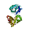

 Mass: 22.990 Da / Num. of mol.: 1 / Source method: obtained synthetically / Formula: Na
 Mass: 90.121 Da / Num. of mol.: 2 / Source method: obtained synthetically / Formula: C4H10O2
 Mass: 18.015 Da / Num. of mol.: 107 / Source method: isolated from a natural source / Formula: H2O
 Sample preparation
 / Beamline: BL11-1 / Wavelength: 0.979224, 0.918370, 0.978940
 Processing