 Movie
Movie Controller

[English] 日本語
 Yorodumi
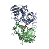
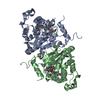
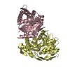
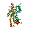
- PDB-2gju: Crystal structure of hypothetical protein PH1004 from Pyrococcus ... -
+ Open data

- Basic information
| Entry | Database: PDB / ID: 2gju | ||||||
|---|---|---|---|---|---|---|---|
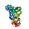
| Title | Crystal structure of hypothetical protein PH1004 from Pyrococcus horikoshii OT3 | ||||||
 Components Components | 252aa long hypothetical protein | ||||||
 Keywords Keywords | STRUCTURAL GENOMICS / UNKNOWN FUNCTION / BETA BARREL / NPPSFA / National Project on Protein Structural and Functional Analyses / RIKEN Structural Genomics/Proteomics Initiative / RSGI | ||||||
| Function / homology | Phosphoesterase MJ0912 / Metallo-dependent phosphatases / Purple Acid Phosphatase; chain A, domain 2 / Metallo-dependent phosphatase-like / 4-Layer Sandwich / Alpha Beta / PHOSPHATE ION / Calcineurin-like phosphoesterase domain-containing protein Function and homology information Function and homology information | ||||||
| Biological species |   Pyrococcus horikoshii (archaea) Pyrococcus horikoshii (archaea) | ||||||
| Method |  X-RAY DIFFRACTION / SYNCHROTRON / MOLECULAR REPLACEMENT / Resolution: 2 Å X-RAY DIFFRACTION / SYNCHROTRON / MOLECULAR REPLACEMENT / Resolution: 2 Å | ||||||
 Authors Authors | Yamamoto, H. / Kunishima, N. / RIKEN Structural Genomics/Proteomics Initiative (RSGI) | ||||||
 Citation Citation | Journal: To be published Title: Crystal structure of hypothetical protein PH1004 from Pyrococcus horikoshii OT3 Authors: Yamamoto, H. / Kunishima, N. | ||||||
| History |
|
- Structure visualization
| Structure viewer | Molecule: MolmilJmol/JSmol |
|---|
- Downloads & links
-Download
| PDBx/mmCIF format | 2gju.cif.gz | 233.6 KB | Display | PDBx/mmCIF format |
|---|---|---|---|---|
| PDB format | pdb2gju.ent.gz | 186.1 KB | Display | PDB format |
| PDBx/mmJSON format | 2gju.json.gz | Tree view | PDBx/mmJSON format | |
| Others |  Other downloads Other downloads |
-Validation report
| Arichive directory | https://data.pdbj.org/pub/pdb/validation_reports/gj/2gjuftp://data.pdbj.org/pub/pdb/validation_reports/gj/2gju | HTTPS FTP |
|---|
-Related structure data
| Related structure data |  1nnwS S: Starting model for refinement |
|---|---|
| Similar structure data | |
| Other databases |
-Links
 PDBj
PDBj
- Assembly
| Deposited unit | 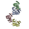
| |||||||||
|---|---|---|---|---|---|---|---|---|---|---|
| 1 | 
| |||||||||
| 2 | 
| |||||||||
| 3 | 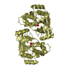
| |||||||||
| Unit cell |
| |||||||||
| Components on special symmetry positions |
| |||||||||
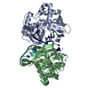
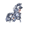
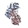
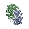
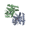
| Details | Biological assembly is dimer in the asymmetric unit. |
-Components
| #1: Protein | Mass: 28678.295 Da / Num. of mol.: 4 Source method: isolated from a genetically manipulated source Source: (gene. exp.) Pyrococcus horikoshii (archaea) / Strain: OT3 / Plasmid: pET11a / Production host:  #2: Chemical | ChemComp-NA /   Mass: 22.990 Da / Num. of mol.: 4 / Source method: obtained synthetically / Formula: Na Mass: 22.990 Da / Num. of mol.: 4 / Source method: obtained synthetically / Formula: Na#3: Chemical | ChemComp-PO4 /   Mass: 94.971 Da / Num. of mol.: 7 / Source method: obtained synthetically / Formula: PO4 Mass: 94.971 Da / Num. of mol.: 7 / Source method: obtained synthetically / Formula: PO4#4: Water | ChemComp-HOH / |  Mass: 18.015 Da / Num. of mol.: 1039 / Source method: isolated from a natural source / Formula: H2O Mass: 18.015 Da / Num. of mol.: 1039 / Source method: isolated from a natural source / Formula: H2O |
|---|
-Experimental details
-Experiment
| Experiment | Method: X-RAY DIFFRACTION / Number of used crystals: 1 |
|---|
- Sample preparation
| Crystal | Density Matthews: 3.1 Å3/Da / Density % sol: 60.26 % |
|---|---|
| Crystal grow | Temperature: 291 K / Method: microbatch Details: 0.05M Potassium dihydrogen phosphate, 10% PEG 8000, MICROBATCH, temperature 291K |
-Data collection
| Diffraction | Mean temperature: 103 K |
|---|---|
| Diffraction source | Source: SYNCHROTRON / Site: SPring-8  / Beamline: BL26B1 / Wavelength: 1 Å / Beamline: BL26B1 / Wavelength: 1 Å |
| Detector | Type: RIGAKU JUPITER 210 / Detector: CCD / Date: Dec 18, 2005 / Details: MIRRORS |
| Radiation | Monochromator: MIRRORS / Protocol: SINGLE WAVELENGTH / Monochromatic (M) / Laue (L): M / Scattering type: x-ray |
| Radiation wavelength | Wavelength: 1 Å / Relative weight: 1 |
| Reflection | Resolution: 2→40 Å / Num. all: 93428 / Num. obs: 91840 / % possible obs: 98.3 % / Observed criterion σ(F): 0 / Observed criterion σ(I): 0 / Redundancy: 4 % / Biso Wilson estimate: 14.3 Å2 / Rmerge(I) obs: 0.095 / Rsym value: 0.086 / Net I/σ(I): 8.5 |
| Reflection shell | Resolution: 2→2.07 Å / Redundancy: 3.7 % / Rmerge(I) obs: 0.281 / Mean I/σ(I) obs: 3.6 / Num. unique all: 8949 / Rsym value: 0.256 / % possible all: 95.7 |
- Processing
| Software |
| |||||||||||||||||||||||||
|---|---|---|---|---|---|---|---|---|---|---|---|---|---|---|---|---|---|---|---|---|---|---|---|---|---|---|
| Refinement | Method to determine structure: MOLECULAR REPLACEMENT Starting model: 1NNW Resolution: 2→39.43 Å / Rfactor Rfree error: 0.003 / Data cutoff high absF: 3998810.95 / Data cutoff low absF: 0 / Isotropic thermal model: RESTRAINED / Cross valid method: THROUGHOUT / σ(F): 0 / σ(I): 0 / Stereochemistry target values: Engh & Huber
| |||||||||||||||||||||||||
| Solvent computation | Solvent model: FLAT MODEL / Bsol: 52.6816 Å2 / ksol: 0.348221 e/Å3 | |||||||||||||||||||||||||
| Displacement parameters | Biso mean: 25.9 Å2
| |||||||||||||||||||||||||
| Refine analyze |
| |||||||||||||||||||||||||
| Refinement step | Cycle: LAST / Resolution: 2→39.43 Å
| |||||||||||||||||||||||||
| Refine LS restraints |
| |||||||||||||||||||||||||
| LS refinement shell | Resolution: 2→2.13 Å / Rfactor Rfree error: 0.01 / Total num. of bins used: 6
| |||||||||||||||||||||||||
| Xplor file |
|
