 Movie
Movie Controller

[English] 日本語
 Yorodumi
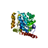
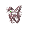
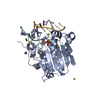
- PDB-2gjn: crystal structure of 2-nitropropane dioxygenase complexed with FM... -
+ Open data

- Basic information
| Entry | Database: PDB / ID: 2gjn | ||||||
|---|---|---|---|---|---|---|---|
| Title | crystal structure of 2-nitropropane dioxygenase complexed with FMN and substrate | ||||||
 Components Components | hypothetical protein PA1024 | ||||||
 Keywords Keywords | OXIDOREDUCTASE / 2-nitropropane dioxygenase / FMN / 2-nitropropane / substrate | ||||||
| Function / homology |  Function and homology information Function and homology informationNADH:quinone reductase (non-electrogenic) / nitronate monooxygenase activity / NADH dehydrogenase (quinone) (non-electrogenic) activity / oxidoreductase activity / nucleotide binding Similarity search - Function | ||||||
| Biological species |  Pseudomonas aeruginosa PAO1 (bacteria) Pseudomonas aeruginosa PAO1 (bacteria) | ||||||
| Method |  X-RAY DIFFRACTION / SYNCHROTRON / MAD / Resolution: 2.3 Å X-RAY DIFFRACTION / SYNCHROTRON / MAD / Resolution: 2.3 Å | ||||||
 Authors Authors | Suh, S.W. | ||||||
 Citation Citation | Journal: J.Biol.Chem. / Year: 2006 Title: Crystal Structure of 2-Nitropropane Dioxygenase Complexed with FMN and Substrate: identification of the catalytic base Authors: Ha, J.Y. / Min, J.Y. / Lee, S.K. / Kim, H.S. / Kim, J. / Kim, K.H. / Lee, H.H. / Kim, H.K. / Yoon, H.J. / Suh, S.W. | ||||||
| History |
|
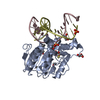
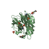
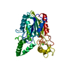
- Structure visualization
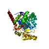
| Structure viewer | Molecule: MolmilJmol/JSmol |
|---|
- Downloads & links
-Download
| PDBx/mmCIF format | 2gjn.cif.gz | 81.2 KB | Display | PDBx/mmCIF format |
|---|---|---|---|---|
| PDB format | pdb2gjn.ent.gz | 59.1 KB | Display | PDB format |
| PDBx/mmJSON format | 2gjn.json.gz | Tree view | PDBx/mmJSON format | |
| Others |  Other downloads Other downloads |
-Validation report
| Arichive directory | https://data.pdbj.org/pub/pdb/validation_reports/gj/2gjnftp://data.pdbj.org/pub/pdb/validation_reports/gj/2gjn | HTTPS FTP |
|---|
-Related structure data


-Links
 PDBj
PDBj- Assembly
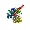
| Deposited unit | 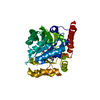
| ||||||||
|---|---|---|---|---|---|---|---|---|---|
| 1 |
| ||||||||
| 2 | 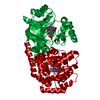
| ||||||||
| Unit cell |
|
-Components
| #1: Protein | Mass: 34860.902 Da / Num. of mol.: 1 Source method: isolated from a genetically manipulated source Source: (gene. exp.) Pseudomonas aeruginosa PAO1 (bacteria) / Species: Pseudomonas aeruginosa / Strain: PA01 / Plasmid: pET21a(+) / Production host: |
|---|---|
| #2: Chemical | ChemComp-FMN /   Mass: 456.344 Da / Num. of mol.: 1 / Source method: obtained synthetically / Formula: C17H21N4O9P Mass: 456.344 Da / Num. of mol.: 1 / Source method: obtained synthetically / Formula: C17H21N4O9P |
| #3: Chemical | ChemComp-NIS /   Mass: 89.093 Da / Num. of mol.: 1 / Source method: obtained synthetically / Formula: C3H7NO2 Mass: 89.093 Da / Num. of mol.: 1 / Source method: obtained synthetically / Formula: C3H7NO2 |
| #4: Water | ChemComp-HOH /  Mass: 18.015 Da / Num. of mol.: 224 / Source method: isolated from a natural source / Formula: H2O Mass: 18.015 Da / Num. of mol.: 224 / Source method: isolated from a natural source / Formula: H2O |
-Experimental details
-Experiment
| Experiment | Method: X-RAY DIFFRACTION / Number of used crystals: 1 |
|---|
- Sample preparation
| Crystal | Density Matthews: 2.88 Å3/Da / Density % sol: 57.34 % |
|---|---|
| Crystal grow | Temperature: 297 K / Method: vapor diffusion, hanging drop / pH: 7 Details: 0.1M HEPES at pH 7.0, 5%(w/v) tacsimate at pH 7.0, 10%(w/v) PEG MME 5000, VAPOR DIFFUSION, HANGING DROP, temperature 297K |
-Data collection
| Diffraction |
| |||||||||||||||||||||||||||||||||||||||||||||||||||||||||||||||||||||||||||||||||||||||||||||||||||||||||||||||||||||||||||||||||||||||||||||||||||||||||||||||||||||||||||||||||||||||||||||||||||||||||||||||||||||||||
|---|---|---|---|---|---|---|---|---|---|---|---|---|---|---|---|---|---|---|---|---|---|---|---|---|---|---|---|---|---|---|---|---|---|---|---|---|---|---|---|---|---|---|---|---|---|---|---|---|---|---|---|---|---|---|---|---|---|---|---|---|---|---|---|---|---|---|---|---|---|---|---|---|---|---|---|---|---|---|---|---|---|---|---|---|---|---|---|---|---|---|---|---|---|---|---|---|---|---|---|---|---|---|---|---|---|---|---|---|---|---|---|---|---|---|---|---|---|---|---|---|---|---|---|---|---|---|---|---|---|---|---|---|---|---|---|---|---|---|---|---|---|---|---|---|---|---|---|---|---|---|---|---|---|---|---|---|---|---|---|---|---|---|---|---|---|---|---|---|---|---|---|---|---|---|---|---|---|---|---|---|---|---|---|---|---|---|---|---|---|---|---|---|---|---|---|---|---|---|---|---|---|---|---|---|---|---|---|---|---|---|---|---|---|---|---|---|---|---|
| Diffraction source | Source: SYNCHROTRON / Site: Photon Factory  / Beamline: AR-NW12A / Wavelength: 0.9791, 0.9749, 0.9500 / Beamline: AR-NW12A / Wavelength: 0.9791, 0.9749, 0.9500 | |||||||||||||||||||||||||||||||||||||||||||||||||||||||||||||||||||||||||||||||||||||||||||||||||||||||||||||||||||||||||||||||||||||||||||||||||||||||||||||||||||||||||||||||||||||||||||||||||||||||||||||||||||||||||
| Detector | Type: ADSC QUANTUM 210 / Detector: CCD / Date: Dec 3, 2003 / Details: mirrors | |||||||||||||||||||||||||||||||||||||||||||||||||||||||||||||||||||||||||||||||||||||||||||||||||||||||||||||||||||||||||||||||||||||||||||||||||||||||||||||||||||||||||||||||||||||||||||||||||||||||||||||||||||||||||
| Radiation | Monochromator: Numerical link type Si(111) double crystal monochromator, liquid nitrogen cooling Protocol: MAD / Monochromatic (M) / Laue (L): M / Scattering type: x-ray | |||||||||||||||||||||||||||||||||||||||||||||||||||||||||||||||||||||||||||||||||||||||||||||||||||||||||||||||||||||||||||||||||||||||||||||||||||||||||||||||||||||||||||||||||||||||||||||||||||||||||||||||||||||||||
| Radiation wavelength |
| |||||||||||||||||||||||||||||||||||||||||||||||||||||||||||||||||||||||||||||||||||||||||||||||||||||||||||||||||||||||||||||||||||||||||||||||||||||||||||||||||||||||||||||||||||||||||||||||||||||||||||||||||||||||||
| Reflection | D res low: 50 Å
| |||||||||||||||||||||||||||||||||||||||||||||||||||||||||||||||||||||||||||||||||||||||||||||||||||||||||||||||||||||||||||||||||||||||||||||||||||||||||||||||||||||||||||||||||||||||||||||||||||||||||||||||||||||||||
| Diffraction reflection shell |
| |||||||||||||||||||||||||||||||||||||||||||||||||||||||||||||||||||||||||||||||||||||||||||||||||||||||||||||||||||||||||||||||||||||||||||||||||||||||||||||||||||||||||||||||||||||||||||||||||||||||||||||||||||||||||
| Reflection | Resolution: 2.2→30 Å / Num. obs: 20734 / % possible obs: 99 % / Rmerge(I) obs: 0.077 / Χ2: 1.113 / Net I/σ(I): 11 | |||||||||||||||||||||||||||||||||||||||||||||||||||||||||||||||||||||||||||||||||||||||||||||||||||||||||||||||||||||||||||||||||||||||||||||||||||||||||||||||||||||||||||||||||||||||||||||||||||||||||||||||||||||||||
| Reflection shell |
|
-Phasing
| Phasing | Method: MAD | ||||||||||||||||||||||||||||||||||||||||||||||||||||||||
|---|---|---|---|---|---|---|---|---|---|---|---|---|---|---|---|---|---|---|---|---|---|---|---|---|---|---|---|---|---|---|---|---|---|---|---|---|---|---|---|---|---|---|---|---|---|---|---|---|---|---|---|---|---|---|---|---|---|
| Phasing set |
| ||||||||||||||||||||||||||||||||||||||||||||||||||||||||
| Phasing MAD | D res high: 2.5 Å / D res low: 20 Å / FOM : 0.68 / Reflection: 14435 | ||||||||||||||||||||||||||||||||||||||||||||||||||||||||
| Phasing MAD set |
| ||||||||||||||||||||||||||||||||||||||||||||||||||||||||
| Phasing MAD set site |
| ||||||||||||||||||||||||||||||||||||||||||||||||||||||||
| Phasing MAD shell |
| ||||||||||||||||||||||||||||||||||||||||||||||||||||||||
| Phasing dm | FOM : 0.73 / FOM acentric: 0.74 / FOM centric: 0.67 / Reflection: 14409 / Reflection acentric: 12813 / Reflection centric: 1596 | ||||||||||||||||||||||||||||||||||||||||||||||||||||||||
| Phasing dm shell |
|
- Processing
| Software |
| ||||||||||||||||||||||||||||||||||||
|---|---|---|---|---|---|---|---|---|---|---|---|---|---|---|---|---|---|---|---|---|---|---|---|---|---|---|---|---|---|---|---|---|---|---|---|---|---|
| Refinement | Method to determine structure: MAD / Resolution: 2.3→15 Å / σ(F): 0
| ||||||||||||||||||||||||||||||||||||
| Solvent computation | Bsol: 68.1 Å2 | ||||||||||||||||||||||||||||||||||||
| Displacement parameters | Biso mean: 28.75 Å2
| ||||||||||||||||||||||||||||||||||||
| Refinement step | Cycle: LAST / Resolution: 2.3→15 Å
| ||||||||||||||||||||||||||||||||||||
| Xplor file |
|
