 Movie
Movie Controller


+ Open data

- Basic information
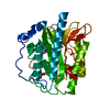
| Entry | Database: PDB / ID: 2ggc | ||||||
|---|---|---|---|---|---|---|---|
| Title | Novel bacterial methionine aminopeptidase inhibitors | ||||||
 Components Components | Methionine aminopeptidase | ||||||
 Keywords Keywords | HYDROLASE / methionine amino peptidase / pita-bread fold / MAP inhibitor / antibacterial | ||||||
| Function / homology |  Function and homology information Function and homology informationmethionyl aminopeptidase / initiator methionyl aminopeptidase activity / metalloaminopeptidase activity / ferrous iron binding / proteolysis / cytosol Similarity search - Function | ||||||
| Biological species |  | ||||||
| Method |  X-RAY DIFFRACTION / SYNCHROTRON / MOLECULAR REPLACEMENT / Resolution: 1 Å X-RAY DIFFRACTION / SYNCHROTRON / MOLECULAR REPLACEMENT / Resolution: 1 Å | ||||||
 Authors Authors | Evdokimov, A.G. / Pokross, M.E. / Walter, R.L. / Mekel, M. | ||||||
 Citation Citation | Journal: Proteins / Year: 2007 Title: Serendipitous discovery of novel bacterial methionine aminopeptidase inhibitors. Authors: Evdokimov, A.G. / Pokross, M. / Walter, R.L. / Mekel, M. / Barnett, B.L. / Amburgey, J. / Seibel, W.L. / Soper, S.J. / Djung, J.F. / Fairweather, N. / Diven, C. / Rastogi, V. / Grinius, L. / ...Authors: Evdokimov, A.G. / Pokross, M. / Walter, R.L. / Mekel, M. / Barnett, B.L. / Amburgey, J. / Seibel, W.L. / Soper, S.J. / Djung, J.F. / Fairweather, N. / Diven, C. / Rastogi, V. / Grinius, L. / Klanke, C. / Siehnel, R. / Twinem, T. / Andrews, R. / Curnow, A. | ||||||
| History |
|
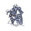
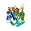
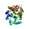
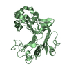
- Structure visualization
| Structure viewer | Molecule: MolmilJmol/JSmol |
|---|
- Downloads & links
-Download
| PDBx/mmCIF format | 2ggc.cif.gz | 140.5 KB | Display | PDBx/mmCIF format |
|---|---|---|---|---|
| PDB format | pdb2ggc.ent.gz | 108.5 KB | Display | PDB format |
| PDBx/mmJSON format | 2ggc.json.gz | Tree view | PDBx/mmJSON format | |
| Others |  Other downloads Other downloads |
-Validation report
| Arichive directory | https://data.pdbj.org/pub/pdb/validation_reports/gg/2ggcftp://data.pdbj.org/pub/pdb/validation_reports/gg/2ggc | HTTPS FTP |
|---|
-Related structure data
| Related structure data |  2gg0SC  2gg2C  2gg3C 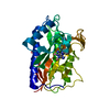 2gg5C 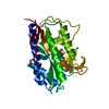 2gg7C  2gg8C  2gg9C 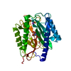 2ggbC S: Starting model for refinement C: citing same article ( |
|---|---|
| Similar structure data |

-Links
 PDBj
PDBj
- Assembly
| Deposited unit | 
| ||||||||
|---|---|---|---|---|---|---|---|---|---|
| 1 |
| ||||||||
| Unit cell |
|
-Components
| #1: Protein | Mass: 29239.643 Da / Num. of mol.: 1 Source method: isolated from a genetically manipulated source Source: (gene. exp.) | ||||||
|---|---|---|---|---|---|---|---|
| #2: Chemical |   Mass: 58.933 Da / Num. of mol.: 2 / Source method: obtained synthetically / Formula: Co Mass: 58.933 Da / Num. of mol.: 2 / Source method: obtained synthetically / Formula: Co#3: Chemical | ChemComp-NA / |   Mass: 22.990 Da / Num. of mol.: 1 / Source method: obtained synthetically / Formula: Na Mass: 22.990 Da / Num. of mol.: 1 / Source method: obtained synthetically / Formula: Na#4: Chemical | ChemComp-MET / |   Type: L-peptide linking / Mass: 149.211 Da / Num. of mol.: 1 / Source method: obtained synthetically / Formula: C5H11NO2S Type: L-peptide linking / Mass: 149.211 Da / Num. of mol.: 1 / Source method: obtained synthetically / Formula: C5H11NO2S#5: Water | ChemComp-HOH / |  Mass: 18.015 Da / Num. of mol.: 345 / Source method: isolated from a natural source / Formula: H2O Mass: 18.015 Da / Num. of mol.: 345 / Source method: isolated from a natural source / Formula: H2O |
-Experimental details
-Experiment
| Experiment | Method: X-RAY DIFFRACTION / Number of used crystals: 1 |
|---|
- Sample preparation
| Crystal | Density Matthews: 1.87 Å3/Da / Density % sol: 34.39 % |
|---|---|
| Crystal grow | Temperature: 298 K / Method: batch / pH: 7 Details: 10 mg/ml protein, 25% PEG 8000, 100 mM TRIS-HCl, 1-5 mM inhibitor, pH 7.0, batch, temperature 298K |
-Data collection
| Diffraction | Mean temperature: 100 K |
|---|---|
| Diffraction source | Source: SYNCHROTRON / Site: APS  / Beamline: 17-ID / Wavelength: 1 Å / Beamline: 17-ID / Wavelength: 1 Å |
| Detector | Type: ADSC QUANTUM 4 / Detector: CCD / Date: Jan 1, 2002 |
| Radiation | Monochromator: Si / Protocol: SINGLE WAVELENGTH / Monochromatic (M) / Laue (L): M / Scattering type: x-ray |
| Radiation wavelength | Wavelength: 1 Å / Relative weight: 1 |
| Reflection | Resolution: 1→23.8 Å / Num. all: 98330 / Num. obs: 98330 / % possible obs: 93.2 % / Observed criterion σ(F): 0 / Observed criterion σ(I): 0 / Redundancy: 2.6 % / Rmerge(I) obs: 0.021 / Net I/σ(I): 28.1 |
| Reflection shell | Resolution: 1→1.15 Å / Redundancy: 1.5 % / Rmerge(I) obs: 0.082 / Mean I/σ(I) obs: 11.2 / Num. unique all: 17489 / % possible all: 73.2 |
- Processing
| Software |
| ||||||||||||||||||||||||||||||||||||||||||||||||||||||||||||||||||||||||||||||||||||||||||||||||||||||||||||||||||||||||||||||||||||||||||||||||||||||
|---|---|---|---|---|---|---|---|---|---|---|---|---|---|---|---|---|---|---|---|---|---|---|---|---|---|---|---|---|---|---|---|---|---|---|---|---|---|---|---|---|---|---|---|---|---|---|---|---|---|---|---|---|---|---|---|---|---|---|---|---|---|---|---|---|---|---|---|---|---|---|---|---|---|---|---|---|---|---|---|---|---|---|---|---|---|---|---|---|---|---|---|---|---|---|---|---|---|---|---|---|---|---|---|---|---|---|---|---|---|---|---|---|---|---|---|---|---|---|---|---|---|---|---|---|---|---|---|---|---|---|---|---|---|---|---|---|---|---|---|---|---|---|---|---|---|---|---|---|---|---|---|
| Refinement | Method to determine structure: MOLECULAR REPLACEMENT Starting model: PDB ENTRY 2GG0 Resolution: 1→23.8 Å / Cor.coef. Fo:Fc: 0.977 / Cor.coef. Fo:Fc free: 0.975 / SU B: 0.498 / SU ML: 0.012 / Cross valid method: THROUGHOUT / σ(F): 0 / σ(I): 0 / ESU R: 0.025 / ESU R Free: 0.024 / Stereochemistry target values: MAXIMUM LIKELIHOOD / Details: HYDROGENS HAVE BEEN ADDED IN THE RIDING POSITIONS
| ||||||||||||||||||||||||||||||||||||||||||||||||||||||||||||||||||||||||||||||||||||||||||||||||||||||||||||||||||||||||||||||||||||||||||||||||||||||
| Solvent computation | Ion probe radii: 0.8 Å / Shrinkage radii: 0.8 Å / VDW probe radii: 1.2 Å / Solvent model: MASK | ||||||||||||||||||||||||||||||||||||||||||||||||||||||||||||||||||||||||||||||||||||||||||||||||||||||||||||||||||||||||||||||||||||||||||||||||||||||
| Displacement parameters | Biso mean: 8.287 Å2
| ||||||||||||||||||||||||||||||||||||||||||||||||||||||||||||||||||||||||||||||||||||||||||||||||||||||||||||||||||||||||||||||||||||||||||||||||||||||
| Refinement step | Cycle: LAST / Resolution: 1→23.8 Å
| ||||||||||||||||||||||||||||||||||||||||||||||||||||||||||||||||||||||||||||||||||||||||||||||||||||||||||||||||||||||||||||||||||||||||||||||||||||||
| Refine LS restraints |
| ||||||||||||||||||||||||||||||||||||||||||||||||||||||||||||||||||||||||||||||||||||||||||||||||||||||||||||||||||||||||||||||||||||||||||||||||||||||
| LS refinement shell | Resolution: 1→1.026 Å / Total num. of bins used: 20
|
