 Movie
Movie Controller

[English] 日本語
 Yorodumi
- PDB-2gc7: Substrate reduced, copper free complex of methylamine dehydrogena... -
+ Open data

- Basic information
| Entry | Database: PDB / ID: 2gc7 | |||||||||
|---|---|---|---|---|---|---|---|---|---|---|
| Title | Substrate reduced, copper free complex of methylamine dehydrogenase, amicyanin and cytochrome c551i from Paracoccus denitrificans. | |||||||||
 Components Components |
| |||||||||
 Keywords Keywords | Oxidoreductase / Electron transport / Electron transfer / Methylamine dehydrogenase / Cytochrome / amicyanin | |||||||||
| Function / homology |  Function and homology information Function and homology informationmethylamine dehydrogenase (amicyanin) / methanol metabolic process / methylamine dehydrogenase (amicyanin) activity / methylamine metabolic process / aliphatic amine dehydrogenase activity / amine metabolic process / outer membrane-bounded periplasmic space / periplasmic space / electron transfer activity / iron ion binding ...methylamine dehydrogenase (amicyanin) / methanol metabolic process / methylamine dehydrogenase (amicyanin) activity / methylamine metabolic process / aliphatic amine dehydrogenase activity / amine metabolic process / outer membrane-bounded periplasmic space / periplasmic space / electron transfer activity / iron ion binding / copper ion binding / heme binding Similarity search - Function | |||||||||
| Biological species |  Paracoccus denitrificans (bacteria) Paracoccus denitrificans (bacteria) | |||||||||
| Method |  X-RAY DIFFRACTION / SYNCHROTRON / Refined directly / Resolution: 1.9 Å X-RAY DIFFRACTION / SYNCHROTRON / Refined directly / Resolution: 1.9 Å | |||||||||
 Authors Authors | Chen, Z. / Durley, R. / Davidson, V.L. / Mathews, F.S. | |||||||||
 Citation Citation | Journal: To be Published Title: Structral comparison of the oxidized ternary electron transfer complex of methylamine dehydrogenase, amicyanin and cytochrome c551i from Paracoccus denitrificans with the substrate-reduced, ...Title: Structral comparison of the oxidized ternary electron transfer complex of methylamine dehydrogenase, amicyanin and cytochrome c551i from Paracoccus denitrificans with the substrate-reduced, copper free complex at 1.9 A resolution. Authors: Chen, Z. / Durley, R. / Davidson, V.L. / Mathews, F.S. #1: Journal: Science / Year: 1994Title: Structure of an electron transfer complex: methylamine dehydrogenase, amicyanin and cytochrome c551i. Authors: Chen, L. / Durley, R. / Mathews, F.S. / Davidson, V.L. | |||||||||
| History |
|
- Structure visualization
| Structure viewer | Molecule: MolmilJmol/JSmol |
|---|
- Downloads & links
-Download
| PDBx/mmCIF format | 2gc7.cif.gz | 611.3 KB | Display | PDBx/mmCIF format |
|---|---|---|---|---|
| PDB format | pdb2gc7.ent.gz | 498.5 KB | Display | PDB format |
| PDBx/mmJSON format | 2gc7.json.gz | Tree view | PDBx/mmJSON format | |
| Others |  Other downloads Other downloads |
-Validation report
| Summary document | 2gc7_validation.pdf.gz | 1.9 MB | Display | wwPDB validaton report |
|---|---|---|---|---|
| Full document | 2gc7_full_validation.pdf.gz | 2 MB | Display | |
| Data in XML | 2gc7_validation.xml.gz | 119.3 KB | Display | |
| Data in CIF | 2gc7_validation.cif.gz | 165.4 KB | Display | |
| Arichive directory | https://data.pdbj.org/pub/pdb/validation_reports/gc/2gc7ftp://data.pdbj.org/pub/pdb/validation_reports/gc/2gc7 | HTTPS FTP |
-Related structure data
| Related structure data |  2gc4S S: Starting model for refinement |
|---|---|
| Similar structure data |


-Links
 PDBj
PDBj


- Assembly
| Deposited unit | 
| ||||||||
|---|---|---|---|---|---|---|---|---|---|
| 1 | 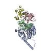
| ||||||||
| 2 | 
| ||||||||
| 3 | 
| ||||||||
| 4 | 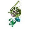
| ||||||||
| Unit cell |
|
-Components
-Protein , 2 types, 8 molecules CGKODHLP
| #3: Protein | Mass: 11505.171 Da / Num. of mol.: 4 / Source method: isolated from a natural source / Source: (natural) Paracoccus denitrificans (bacteria) / References: UniProt: P22364#4: Protein | Mass: 16274.852 Da / Num. of mol.: 4 / Source method: isolated from a natural source / Source: (natural) Paracoccus denitrificans (bacteria) / References: UniProt: P29899 |
|---|
-Methylamine dehydrogenase ... / Antibody , 2 types, 8 molecules AEIMBFJN
| #1: Protein | Mass: 42449.277 Da / Num. of mol.: 4 / Source method: isolated from a natural source / Source: (natural) Paracoccus denitrificans (bacteria)References: GenBank: 69934851, UniProt: P29894*PLUS, EC: 1.4.99.3 #2: Antibody | Mass: 14210.696 Da / Num. of mol.: 4 / Source method: isolated from a natural source / Source: (natural) Paracoccus denitrificans (bacteria) / References: UniProt: P22619, EC: 1.4.99.3 |
|---|
-Non-polymers , 3 types, 1242 molecules 


| #5: Chemical | ChemComp-NA /  Mass: 22.990 Da / Num. of mol.: 4 / Source method: obtained synthetically / Formula: Na Mass: 22.990 Da / Num. of mol.: 4 / Source method: obtained synthetically / Formula: Na#6: Chemical | ChemComp-HEC /  Mass: 618.503 Da / Num. of mol.: 4 / Source method: obtained synthetically / Formula: C34H34FeN4O4 Mass: 618.503 Da / Num. of mol.: 4 / Source method: obtained synthetically / Formula: C34H34FeN4O4#7: Water | ChemComp-HOH / | Mass: 18.015 Da / Num. of mol.: 1234 / Source method: isolated from a natural source / Formula: H2O |
|---|
-Experimental details
-Experiment
| Experiment | Method: X-RAY DIFFRACTION / Number of used crystals: 1 |
|---|
- Sample preparation
| Crystal | Density Matthews: 2.89 Å3/Da / Density % sol: 57.45 % |
|---|---|
| Crystal grow | Temperature: 295 K / Method: vapor diffusion, sitting drop / pH: 5.5 Details: 2.3-2.6M sodium/potassium phosphate, pH 5.5, VAPOR DIFFUSION, SITTING DROP, temperature 295K |
-Data collection
| Diffraction | Mean temperature: 298 K |
|---|---|
| Diffraction source | Source: SYNCHROTRON / Site: Photon Factory  / Beamline: BL-6A / Wavelength: 0.95 Å / Beamline: BL-6A / Wavelength: 0.95 Å |
| Detector | Type: WEISSENBERG / Detector: DIFFRACTOMETER / Date: May 25, 1996 |
| Radiation | Protocol: SINGLE WAVELENGTH / Monochromatic (M) / Laue (L): M / Scattering type: x-ray |
| Radiation wavelength | Wavelength: 0.95 Å / Relative weight: 1 |
| Reflection | Resolution: 1.9→30 Å / Num. all: 295619 / Num. obs: 254385 / % possible obs: 83.2 % / Observed criterion σ(F): 0 / Observed criterion σ(I): 0 / Redundancy: 1.8 % / Biso Wilson estimate: 17.6 Å2 / Rmerge(I) obs: 0.055 / Net I/σ(I): 12.4 |
| Reflection shell | Resolution: 1.9→1.97 Å / Redundancy: 1.7 % / Rmerge(I) obs: 0.276 / Mean I/σ(I) obs: 2.2 / Num. unique all: 13233 / % possible all: 44.8 |
- Processing
| Software |
| |||||||||||||||||||||||||||||||||||||||||||||||||
|---|---|---|---|---|---|---|---|---|---|---|---|---|---|---|---|---|---|---|---|---|---|---|---|---|---|---|---|---|---|---|---|---|---|---|---|---|---|---|---|---|---|---|---|---|---|---|---|---|---|---|
| Refinement | Method to determine structure: Refined directly Starting model: PDB entry 2GC4 Resolution: 1.9→29.94 Å / Rfactor Rfree error: 0.001 / Data cutoff high absF: 217447.43 / Data cutoff low absF: 0 / Isotropic thermal model: Isotropic / Cross valid method: THROUGHOUT / σ(F): 0 / σ(I): 0 / Stereochemistry target values: Engh & Huber
| |||||||||||||||||||||||||||||||||||||||||||||||||
| Displacement parameters | Biso mean: 31.7 Å2 | |||||||||||||||||||||||||||||||||||||||||||||||||
| Refine analyze |
| |||||||||||||||||||||||||||||||||||||||||||||||||
| Refinement step | Cycle: LAST / Resolution: 1.9→29.94 Å
| |||||||||||||||||||||||||||||||||||||||||||||||||
| Refine LS restraints |
| |||||||||||||||||||||||||||||||||||||||||||||||||
| LS refinement shell | Refine-ID: X-RAY DIFFRACTION / Total num. of bins used: 6
|
