Mass: 18.015 Da / Num. of mol.: 193 / Source method: isolated from a natural source / Formula: H2O
Has protein modification
Y
-
Experimental details
-
Experiment
Experiment
Method: X-RAY DIFFRACTION
-
Sample preparation
Crystal
Density Matthews: 2.64 Å3/Da / Density % sol: 53.37 %
Crystal grow
Temperature: 298 K / Method: vapor diffusion, hanging drop / pH: 4.2 Details: Well solution: 1.5-1.6 M ammonium sulfate, 0.1 M citrate/phosphate buffer pH 4.2. Protein solution: 5 mg/ml in 0.025 M Tris ph 7.5, 0.0005 M EDTA, 0.0005 M DTT, 0.1 M sodium chloride. Drops: ...Details: Well solution: 1.5-1.6 M ammonium sulfate, 0.1 M citrate/phosphate buffer pH 4.2. Protein solution: 5 mg/ml in 0.025 M Tris ph 7.5, 0.0005 M EDTA, 0.0005 M DTT, 0.1 M sodium chloride. Drops: 1:1 well:protein., VAPOR DIFFUSION, HANGING DROP, temperature 298K
Protocol: SINGLE WAVELENGTH / Monochromatic (M) / Laue (L): M / Scattering type: x-ray
Radiation wavelength
Wavelength: 0.979 Å / Relative weight: 1
Reflection
Redundancy: 13 % / Av σ(I) over netI: 8.2 / Number: 566822 / Rmerge(I) obs: 0.113 / Χ2: 1.99 / D res high: 2.1 Å / D res low: 50 Å / Num. obs: 43510 / % possible obs: 99.4
Diffraction reflection shell
ID: 1
Highest resolution (Å)
Lowest resolution (Å)
Num. obs
% possible obs (%)
Rmerge(I) obs
Chi squared
Redundancy
4.52
50
4591
99.9
0.082
5.089
13.2
3.59
4.52
4417
100
0.08
3.456
14
3.14
3.59
4375
100
0.097
2.613
14.3
2.85
3.14
4380
100
0.127
1.847
14.4
2.65
2.85
4341
100
0.181
1.351
14.5
2.49
2.65
4339
100
0.23
1.13
14.5
2.37
2.49
4356
100
0.289
0.971
14.1
2.26
2.37
4321
99.9
0.35
0.909
12.4
2.18
2.26
4284
99.1
0.42
0.773
10.4
2.1
2.18
4106
95.3
0.469
0.717
8
Reflection
Resolution: 2.1→50 Å / Num. obs: 43510 / % possible obs: 99.4 % / Redundancy: 13 % / Rmerge(I) obs: 0.113 / Χ2: 1.991 / Net I/σ(I): 8.2
In the structure databanks used in Yorodumi, some data are registered as the other names, "COVID-19 virus" and "2019-nCoV". Here are the details of the virus and the list of structure data.
Jan 31, 2019. EMDB accession codes are about to change! (news from PDBe EMDB page)
EMDB accession codes are about to change! (news from PDBe EMDB page)
The allocation of 4 digits for EMDB accession codes will soon come to an end. Whilst these codes will remain in use, new EMDB accession codes will include an additional digit and will expand incrementally as the available range of codes is exhausted. The current 4-digit format prefixed with “EMD-” (i.e. EMD-XXXX) will advance to a 5-digit format (i.e. EMD-XXXXX), and so on. It is currently estimated that the 4-digit codes will be depleted around Spring 2019, at which point the 5-digit format will come into force.
The EM Navigator/Yorodumi systems omit the EMD- prefix.
Related info.:Q: What is EMD? / ID/Accession-code notation in Yorodumi/EM Navigator
Yorodumi is a browser for structure data from EMDB, PDB, SASBDB, etc.
This page is also the successor to EM Navigator detail page, and also detail information page/front-end page for Omokage search.
The word "yorodu" (or yorozu) is an old Japanese word meaning "ten thousand". "mi" (miru) is to see.
Related info.:EMDB / PDB / SASBDB / Comparison of 3 databanks / Yorodumi Search / Aug 31, 2016. New EM Navigator & Yorodumi / Yorodumi Papers / Jmol/JSmol / Function and homology information / Changes in new EM Navigator and Yorodumi
 Movie
Movie Controller

 Yorodumi
 Open data
 Basic information
 Components
 Keywords
 Function and homology information
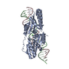
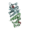
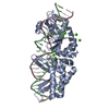
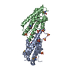
 Yersinia pestis biovar Microtus str. 91001 (bacteria)
 X-RAY DIFFRACTION /
  Authors
 Citation
 Structure visualization
 Downloads & links
 Other downloads


 PDBj
PDBj
 Assembly

 Mass: 96.063 Da / Num. of mol.: 13 / Source method: obtained synthetically / Formula: SO4
 Mass: 192.124 Da / Num. of mol.: 4 / Source method: obtained synthetically / Formula: C6H8O7
 Mass: 18.015 Da / Num. of mol.: 193 / Source method: isolated from a natural source / Formula: H2O
 Sample preparation
 / Beamline: X29A / Wavelength: 0.979 Å
 Processing