Monochromator: Single crystal Si(111) bent monochromator (horizontal focusing) Protocol: SINGLE WAVELENGTH / Monochromatic (M) / Laue (L): M / Scattering type: x-ray
Radiation wavelength
Wavelength: 0.979386 Å / Relative weight: 1
Reflection
Resolution: 2.496→107.211 Å / Num. obs: 75747 / % possible obs: 97.5 % / Redundancy: 3.4 % / Rmerge(I) obs: 0.139 / Rsym value: 0.139 / Net I/σ(I): 3.4
Reflection shell
Diffraction-ID: 1
Resolution (Å)
Redundancy (%)
Rmerge(I) obs
Mean I/σ(I) obs
Num. measured all
Num. unique obs
Rsym value
% possible all
2.5-2.56
1.9
0.2
3.5
9584
5158
0.2
89.4
2.56-2.63
1.9
0.169
4
10013
5290
0.169
96
2.63-2.71
1.9
0.144
4.5
9890
5207
0.144
95.4
2.71-2.79
1.9
0.112
6
9602
5040
0.112
95.5
2.79-2.88
1.9
0.099
7.1
9315
4885
0.099
95.1
2.88-2.98
1.9
0.086
7.5
8906
4672
0.086
94.8
2.98-3.1
4.1
0.447
1.4
19558
4734
0.447
99.4
3.1-3.22
4.5
0.386
1.6
20886
4613
0.386
100
3.22-3.37
4.5
0.297
2.1
19885
4409
0.297
100
3.37-3.53
4.5
0.228
1.6
19151
4233
0.228
100
3.53-3.72
4.5
0.174
2.9
17991
3999
0.174
99.8
3.72-3.95
4.5
0.138
3.9
17198
3803
0.138
99.8
3.95-4.22
4.5
0.114
4.5
15955
3571
0.114
99.9
4.22-4.56
4.5
0.099
5.2
14800
3309
0.099
99.7
4.56-4.99
4.4
0.093
5.1
13628
3069
0.093
99.6
4.99-5.58
4.4
0.098
4.7
11953
2742
0.098
99.7
5.58-6.44
4.2
0.088
5.1
10226
2441
0.088
99.5
6.44-7.89
4.3
0.07
6.6
8917
2069
0.07
99.6
7.89-11.16
4.6
0.052
7.2
7448
1615
0.052
100
11.16-107.21
4.4
0.057
5.3
3894
888
0.057
98.8
-
Phasing
Phasing
Method: molecular replacement
-
Processing
Software
Name
Version
Classification
NB
REFMAC
5.2.0005
refinement
SCALA
datascaling
PDB_EXTRACT
1.701
dataextraction
MOSFLM
datareduction
CCP4
(SCALA)
datascaling
MOLREP
phasing
Refinement
Method to determine structure: MOLECULAR REPLACEMENT Starting model: pdb entry 1xi9A Resolution: 2.5→107.03 Å / Cor.coef. Fo:Fc: 0.945 / Cor.coef. Fo:Fc free: 0.92 / SU B: 27.542 / SU ML: 0.282 / TLS residual ADP flag: LIKELY RESIDUAL / Cross valid method: THROUGHOUT / σ(F): 0 / ESU R Free: 0.347 / Stereochemistry target values: MAXIMUM LIKELIHOOD Details: 1. HYDROGENS HAVE BEEN ADDED IN THE RIDING POSITIONS. 2. ELECTRON DENSITY MAPS INDICATE THAT THE SIDECHAINS OF LYS 232 ON THE SIX SUBUNITS IN THE ASYMMETRIC UNIT FORM A SCHIFF BASE COVALENT ...Details: 1. HYDROGENS HAVE BEEN ADDED IN THE RIDING POSITIONS. 2. ELECTRON DENSITY MAPS INDICATE THAT THE SIDECHAINS OF LYS 232 ON THE SIX SUBUNITS IN THE ASYMMETRIC UNIT FORM A SCHIFF BASE COVALENT BOND WITH THE BOUND PYRIDOXAL PHOSPHATE COFACTOR. 3. A MET-INHIBITION PROTOCOL WAS USED FOR SELENOMETHIONINE INCORPORATION DURING PROTEIN EXPRESSION. THE OCCUPANCY OF THE SE ATOMS IN THE MSE RESIDUES WAS REDUCED TO 0.75 FOR THE REDUCED SCATTERING POWER DUE TO PARTIAL S-MET INCORPORATION. 4. THE CONFORMATION OF GLU 206 IN ALL SIX SUBUNITS IS IS SUPPORTED BY ELECTRON DENSITY EVEN THOUGH THIS RESIDUE IS A RAMACHANDRAN OUTLIER.
Rfactor
Num. reflection
% reflection
Selection details
Rfree
0.266
3781
5 %
RANDOM
Rwork
0.229
-
-
-
obs
0.231
75283
97.52 %
-
Solvent computation
Ion probe radii: 0.8 Å / Shrinkage radii: 0.8 Å / VDW probe radii: 1.2 Å / Solvent model: BABINET MODEL WITH MASK
Displacement parameters
Biso mean: 28.427 Å2
Baniso -1
Baniso -2
Baniso -3
1-
-3.1 Å2
0 Å2
1.06 Å2
2-
-
1.78 Å2
0 Å2
3-
-
-
2.13 Å2
Refinement step
Cycle: LAST / Resolution: 2.5→107.03 Å
Protein
Nucleic acid
Ligand
Solvent
Total
Num. atoms
18179
0
0
190
18369
Refine LS restraints
Refine-ID
Type
Dev ideal
Dev ideal target
Number
X-RAY DIFFRACTION
r_bond_refined_d
0.01
0.022
18624
X-RAY DIFFRACTION
r_bond_other_d
0.002
0.02
17357
X-RAY DIFFRACTION
r_angle_refined_deg
1.526
1.976
25290
X-RAY DIFFRACTION
r_angle_other_deg
0.927
3
39962
X-RAY DIFFRACTION
r_dihedral_angle_1_deg
1.537
5
2350
X-RAY DIFFRACTION
r_dihedral_angle_2_deg
21.265
23.164
787
X-RAY DIFFRACTION
r_dihedral_angle_3_deg
8.558
15
3040
X-RAY DIFFRACTION
r_dihedral_angle_4_deg
8.21
15
132
X-RAY DIFFRACTION
r_chiral_restr
0.091
0.2
2881
X-RAY DIFFRACTION
r_gen_planes_refined
0.004
0.02
20773
X-RAY DIFFRACTION
r_gen_planes_other
0.001
0.02
3939
X-RAY DIFFRACTION
r_nbd_refined
0.189
0.3
3940
X-RAY DIFFRACTION
r_nbd_other
0.153
0.3
18045
X-RAY DIFFRACTION
r_nbtor_refined
0.177
0.5
9489
X-RAY DIFFRACTION
r_nbtor_other
0.085
0.5
10656
X-RAY DIFFRACTION
r_xyhbond_nbd_refined
0.169
0.5
894
X-RAY DIFFRACTION
r_xyhbond_nbd_other
0.03
0.5
5
X-RAY DIFFRACTION
r_symmetry_vdw_refined
0.155
0.3
54
X-RAY DIFFRACTION
r_symmetry_vdw_other
0.175
0.3
154
X-RAY DIFFRACTION
r_symmetry_hbond_refined
0.263
0.5
16
X-RAY DIFFRACTION
r_symmetry_hbond_other
0.07
0.5
1
X-RAY DIFFRACTION
r_mcbond_it
0.508
2
12184
X-RAY DIFFRACTION
r_mcbond_other
0.167
2
4753
X-RAY DIFFRACTION
r_mcangle_it
0.629
4
18834
X-RAY DIFFRACTION
r_scbond_it
1.731
6
7479
X-RAY DIFFRACTION
r_scangle_it
2.472
8
6453
Refine LS restraints NCS
Ens-ID: 1 / Refine-ID: X-RAY DIFFRACTION
Dom-ID
Auth asym-ID
Number
Type
Rms dev position (Å)
Weight position
1
A
2260
TIGHTPOSITIONAL
0.02
0.05
2
B
2260
TIGHTPOSITIONAL
0.02
0.05
3
C
2260
TIGHTPOSITIONAL
0.03
0.05
4
D
2260
TIGHTPOSITIONAL
0.02
0.05
5
E
2260
TIGHTPOSITIONAL
0.02
0.05
6
F
2260
TIGHTPOSITIONAL
0.02
0.05
1
A
3275
MEDIUMPOSITIONAL
0.47
0.5
2
B
3275
MEDIUMPOSITIONAL
0.48
0.5
3
C
3275
MEDIUMPOSITIONAL
0.42
0.5
4
D
3275
MEDIUMPOSITIONAL
0.5
0.5
5
E
3275
MEDIUMPOSITIONAL
0.5
0.5
6
F
3275
MEDIUMPOSITIONAL
0.47
0.5
1
A
2260
TIGHTTHERMAL
0.04
0.5
2
B
2260
TIGHTTHERMAL
0.04
0.5
3
C
2260
TIGHTTHERMAL
0.04
0.5
4
D
2260
TIGHTTHERMAL
0.03
0.5
5
E
2260
TIGHTTHERMAL
0.04
0.5
6
F
2260
TIGHTTHERMAL
0.04
0.5
1
A
3275
MEDIUMTHERMAL
0.26
2
2
B
3275
MEDIUMTHERMAL
0.25
2
3
C
3275
MEDIUMTHERMAL
0.25
2
4
D
3275
MEDIUMTHERMAL
0.22
2
5
E
3275
MEDIUMTHERMAL
0.26
2
6
F
3275
MEDIUMTHERMAL
0.3
2
LS refinement shell
Resolution: 2.5→2.565 Å / Total num. of bins used: 20
Rfactor
Num. reflection
% reflection
Rfree
0.358
249
-
Rwork
0.321
5014
-
obs
-
5263
92.48 %
Refinement TLS params.
Method: refined / Refine-ID: X-RAY DIFFRACTION
ID
L11 (°2)
L12 (°2)
L13 (°2)
L22 (°2)
L23 (°2)
L33 (°2)
S11 (Å °)
S12 (Å °)
S13 (Å °)
S21 (Å °)
S22 (Å °)
S23 (Å °)
S31 (Å °)
S32 (Å °)
S33 (Å °)
T11 (Å2)
T12 (Å2)
T13 (Å2)
T22 (Å2)
T23 (Å2)
T33 (Å2)
Origin x (Å)
Origin y (Å)
Origin z (Å)
1
2.3236
0.0931
-0.7878
2.7765
0.6301
1.6221
-0.0098
0.0153
-0.2046
-0.2468
-0.0041
-0.2627
-0.0001
0.2223
0.0139
0.0018
0.0272
0.0042
-0.0269
-0.0263
-0.0517
-5.633
0.117
15.537
2
3.3299
0.2097
-0.3579
1.6126
0.1528
1.3583
0.1855
-0.0065
0.0208
0.0527
-0.0922
0.2002
-0.1543
-0.3746
-0.0933
0.0161
0.0199
-0.0255
0.099
-0.0728
-0.0877
-37.67
8.808
18.566
3
2.6178
-0.569
-1.0028
2.5988
-0.1182
1.9906
0.0174
0.0702
-0.0386
0.2305
-0.0486
0.2786
-0.1415
-0.3004
0.0312
0.0268
0.0193
-0.0322
-0.0351
-0.0645
-0.0098
-23.729
-36.252
55.666
4
5.0659
0.1658
-1.1337
1.9137
-0.248
1.929
0.3521
0.1897
0.3217
-0.0434
-0.0417
-0.2379
-0.1947
0.3718
-0.3103
-0.0117
-0.0077
-0.0423
0.1515
-0.1113
0.0669
8.479
-27.644
52.589
5
4.1197
-0.0488
-0.5542
1.6265
-0.1421
1.1078
0.2097
-0.0644
0.3329
-0.0207
-0.0594
-0.2264
-0.1612
0.3543
-0.1503
-0.0012
-0.0422
-0.0534
0.0217
-0.0736
-0.0387
8.666
44.148
51.531
6
2.4552
-0.6523
-0.7524
2.7637
-0.2446
1.6781
-0.0251
-0.095
0.0124
0.3207
0.0296
0.2824
-0.04
-0.2048
-0.0044
-0.0242
-0.0187
-0.022
-0.1089
-0.0495
-0.1664
-23.269
35.586
54.425
Refinement TLS group
Refine-ID: X-RAY DIFFRACTION / Selection: ALL
ID
Refine TLS-ID
Auth asym-ID
Label asym-ID
Auth seq-ID
Label seq-ID
1
1
A
A
4 - 392
16 - 404
2
2
B
B
-1 - 392
11 - 404
3
3
C
C
2 - 392
14 - 404
4
4
D
D
1 - 390
13 - 402
5
5
E
E
-1 - 391
11 - 403
6
6
F
F
3 - 391
15 - 403
+
About Yorodumi
-
News
-
Feb 9, 2022. New format data for meta-information of EMDB entries
New format data for meta-information of EMDB entries
Version 3 of the EMDB header file is now the official format.
The previous official version 1.9 will be removed from the archive.
In the structure databanks used in Yorodumi, some data are registered as the other names, "COVID-19 virus" and "2019-nCoV". Here are the details of the virus and the list of structure data.
Jan 31, 2019. EMDB accession codes are about to change! (news from PDBe EMDB page)
EMDB accession codes are about to change! (news from PDBe EMDB page)
The allocation of 4 digits for EMDB accession codes will soon come to an end. Whilst these codes will remain in use, new EMDB accession codes will include an additional digit and will expand incrementally as the available range of codes is exhausted. The current 4-digit format prefixed with “EMD-” (i.e. EMD-XXXX) will advance to a 5-digit format (i.e. EMD-XXXXX), and so on. It is currently estimated that the 4-digit codes will be depleted around Spring 2019, at which point the 5-digit format will come into force.
The EM Navigator/Yorodumi systems omit the EMD- prefix.
Related info.:Q: What is EMD? / ID/Accession-code notation in Yorodumi/EM Navigator
Yorodumi is a browser for structure data from EMDB, PDB, SASBDB, etc.
This page is also the successor to EM Navigator detail page, and also detail information page/front-end page for Omokage search.
The word "yorodu" (or yorozu) is an old Japanese word meaning "ten thousand". "mi" (miru) is to see.
Related info.:EMDB / PDB / SASBDB / Comparison of 3 databanks / Yorodumi Search / Aug 31, 2016. New EM Navigator & Yorodumi / Yorodumi Papers / Jmol/JSmol / Function and homology information / Changes in new EM Navigator and Yorodumi
 Movie
Movie Controller

 Yorodumi
 Open data
 Basic information
 Components
 Keywords
 Function and homology information
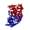
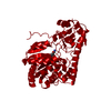
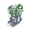
 Thermotoga maritima (bacteria)
 X-RAY DIFFRACTION /
  Authors
 Citation
 Structure visualization
 Downloads & links
 Other downloads
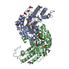


 PDBj
PDBj Assembly

 Mass: 18.015 Da / Num. of mol.: 190 / Source method: isolated from a natural source / Formula: H2O
 Sample preparation
 / Beamline: BL11-1 / Wavelength: 0.979386
  Processing