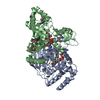
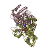
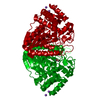
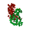
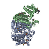
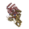
- PDB-2g8y: The structure of a putative malate/lactate dehydrogenase from E. coli. -
+
Open data
ID or keywords:
Loading...
-
Basic information
Entry
Database: PDB / ID: 2g8y
Title
The structure of a putative malate/lactate dehydrogenase from E. coli.
Components
Malate/L-lactate dehydrogenases
Keywords
OXIDOREDUCTASE / malate / lactate / dehydrogenase / NAD / E.coli / Structural Genomics / PSI / Protein Structure Initiative / Midwest Center for Structural Genomics / MCSG
Function / homology
Function and homology information
(R)-4-hydroxyphenyllactate dehydrogenase (NADP+) activity / hydroxyphenylpyruvate reductase / (2R)-hydroxyphenylpyruvate reductase [NAD(P)H] activity / Oxidoreductases; Acting on the CH-OH group of donors; With NAD+ or NADP+ as acceptor / cytosol Similarity search - Function
Malate/L-lactate dehydrogenase-like / Malate/L-sulfolactate/L-lactate dehydrogenase-like superfamily / Malate/L-sulfolactate/L-lactate dehydrogenase-like, NADPH binding domain / Malate/L-sulfolactate/L-lactate dehydrogenase-like, alpha-helical domain / Malate/L-lactate dehydrogenase / Hypothetical Oxidoreductase Yiak; Chain: A, domain 1 / Malate/L-lactate/L-sulpholactate dehydrogenase, four-helix barrel / Malate/L-lactate/L-sulpholactate dehydrogenase, NADPH binding domain / Single helix bin / Ribosomal Protein S8; Chain: A, domain 1 ...Malate/L-lactate dehydrogenase-like / Malate/L-sulfolactate/L-lactate dehydrogenase-like superfamily / Malate/L-sulfolactate/L-lactate dehydrogenase-like, NADPH binding domain / Malate/L-sulfolactate/L-lactate dehydrogenase-like, alpha-helical domain / Malate/L-lactate dehydrogenase / Hypothetical Oxidoreductase Yiak; Chain: A, domain 1 / Malate/L-lactate/L-sulpholactate dehydrogenase, four-helix barrel / Malate/L-lactate/L-sulpholactate dehydrogenase, NADPH binding domain / Single helix bin / Ribosomal Protein S8; Chain: A, domain 1 / Single alpha-helices involved in coiled-coils or other helix-helix interfaces / Up-down Bundle / 2-Layer Sandwich / Orthogonal Bundle / Mainly Alpha / Alpha Beta Similarity search - Domain/homology
SEQUENCE Residue at position -21 is an initiating methionine and a modified residue. Authors ...SEQUENCE Residue at position -21 is an initiating methionine and a modified residue. Authors indicate that the conflict involving residue 53 which is a PHE in the coordinates and ILE in the sequence database reference could be either a sequencing or cloning error
Remark 300
BIOMOLECULE: 1 THIS ENTRY CONTAINS THE CRYSTALLOGRAPHIC ASYMMETRIC UNIT WHICH CONSISTS OF 2 CHAIN(S) ...BIOMOLECULE: 1 THIS ENTRY CONTAINS THE CRYSTALLOGRAPHIC ASYMMETRIC UNIT WHICH CONSISTS OF 2 CHAIN(S). THE BIOLOGICAL UNIT OF THE PROTEIN IS UNKNOWN. PISA SUGGESTS IT TO BE A DIMER WHILE PQS SUGGESTS A TETRAMER
In the structure databanks used in Yorodumi, some data are registered as the other names, "COVID-19 virus" and "2019-nCoV". Here are the details of the virus and the list of structure data.
Jan 31, 2019. EMDB accession codes are about to change! (news from PDBe EMDB page)
EMDB accession codes are about to change! (news from PDBe EMDB page)
The allocation of 4 digits for EMDB accession codes will soon come to an end. Whilst these codes will remain in use, new EMDB accession codes will include an additional digit and will expand incrementally as the available range of codes is exhausted. The current 4-digit format prefixed with “EMD-” (i.e. EMD-XXXX) will advance to a 5-digit format (i.e. EMD-XXXXX), and so on. It is currently estimated that the 4-digit codes will be depleted around Spring 2019, at which point the 5-digit format will come into force.
The EM Navigator/Yorodumi systems omit the EMD- prefix.
Related info.:Q: What is EMD? / ID/Accession-code notation in Yorodumi/EM Navigator
Yorodumi is a browser for structure data from EMDB, PDB, SASBDB, etc.
This page is also the successor to EM Navigator detail page, and also detail information page/front-end page for Omokage search.
The word "yorodu" (or yorozu) is an old Japanese word meaning "ten thousand". "mi" (miru) is to see.
Related info.:EMDB / PDB / SASBDB / Comparison of 3 databanks / Yorodumi Search / Aug 31, 2016. New EM Navigator & Yorodumi / Yorodumi Papers / Jmol/JSmol / Function and homology information / Changes in new EM Navigator and Yorodumi
 Movie
Movie Controller

 Yorodumi
 Open data
 Basic information
 Components
 Keywords
 Function and homology information
 X-RAY DIFFRACTION /
  Authors
 Citation
 Structure visualization
 Downloads & links
 Other downloads
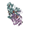

 PDBj
PDBj Assembly
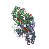


 Mass: 96.063 Da / Num. of mol.: 8 / Source method: obtained synthetically / Formula: SO4
 Mass: 663.425 Da / Num. of mol.: 2 / Source method: obtained synthetically / Formula: C21H27N7O14P2 / Comment: NAD*YM
 Mass: 62.068 Da / Num. of mol.: 10 / Source method: obtained synthetically / Formula: C2H6O2
 Mass: 238.278 Da / Num. of mol.: 1 / Source method: obtained synthetically / Formula: C10H22O6 / Comment: precipitant*YM
 Sample preparation
 / Beamline: 19-ID / Wavelength: 0.97923, 0.97937, 1.00314
 Processing