 Movie
Movie Controller

[English] 日本語
 Yorodumi
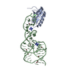
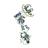
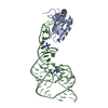
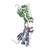
- PDB-2g7c: Clostridium difficile Toxin A Fragment Bound to aGal(1,3)bGal(1,4... -
+ Open data

- Basic information
| Entry | Database: PDB / ID: 2g7c | |||||||||
|---|---|---|---|---|---|---|---|---|---|---|
| Title | Clostridium difficile Toxin A Fragment Bound to aGal(1,3)bGal(1,4)bGlcNAc | |||||||||
 Components Components | Toxin A | |||||||||
 Keywords Keywords | TOXIN / Linear B trisaccharide / protein-carbohydrate complex / bacterial toxin | |||||||||
| Function / homology |  Function and homology information Function and homology informationTransferases; Glycosyltransferases; Hexosyltransferases / host cell cytosol / glycosyltransferase activity / cysteine-type peptidase activity / host cell endosome membrane / toxin activity / Hydrolases; Acting on peptide bonds (peptidases); Cysteine endopeptidases / lipid binding / host cell plasma membrane / proteolysis ...Transferases; Glycosyltransferases; Hexosyltransferases / host cell cytosol / glycosyltransferase activity / cysteine-type peptidase activity / host cell endosome membrane / toxin activity / Hydrolases; Acting on peptide bonds (peptidases); Cysteine endopeptidases / lipid binding / host cell plasma membrane / proteolysis / extracellular region / metal ion binding / membrane Similarity search - Function | |||||||||
| Biological species |  Clostridium difficile (bacteria) Clostridium difficile (bacteria) | |||||||||
| Method |  X-RAY DIFFRACTION / SYNCHROTRON / MOLECULAR REPLACEMENT / Resolution: 2 Å X-RAY DIFFRACTION / SYNCHROTRON / MOLECULAR REPLACEMENT / Resolution: 2 Å | |||||||||
 Authors Authors | Greco, A. / Ho, J.G.S. / Lin, S.J. / Palcic, M.M. / Rupnik, M. / Ng, K.K.S. | |||||||||
 Citation Citation | Journal: Nat.Struct.Mol.Biol. / Year: 2006 Title: Carbohydrate recognition by Clostridium difficile toxin A. Authors: Greco, A. / Ho, J.G. / Lin, S.J. / Palcic, M.M. / Rupnik, M. / Ng, K.K. | |||||||||
| History |
| |||||||||
| Remark 999 | Sequence The differences between the sequence present in this structure and the sequence in the ...Sequence The differences between the sequence present in this structure and the sequence in the reference database are unique to strain 48489 of Clostridium difficile. |
- Structure visualization
| Structure viewer | Molecule: MolmilJmol/JSmol |
|---|
- Downloads & links
-Download
| PDBx/mmCIF format | 2g7c.cif.gz | 125 KB | Display | PDBx/mmCIF format |
|---|---|---|---|---|
| PDB format | pdb2g7c.ent.gz | 96.2 KB | Display | PDB format |
| PDBx/mmJSON format | 2g7c.json.gz | Tree view | PDBx/mmJSON format | |
| Others |  Other downloads Other downloads |
-Validation report
| Summary document | 2g7c_validation.pdf.gz | 1.7 MB | Display | wwPDB validaton report |
|---|---|---|---|---|
| Full document | 2g7c_full_validation.pdf.gz | 1.7 MB | Display | |
| Data in XML | 2g7c_validation.xml.gz | 25.1 KB | Display | |
| Data in CIF | 2g7c_validation.cif.gz | 36.6 KB | Display | |
| Arichive directory | https://data.pdbj.org/pub/pdb/validation_reports/g7/2g7cftp://data.pdbj.org/pub/pdb/validation_reports/g7/2g7c | HTTPS FTP |
-Related structure data
| Related structure data |  2f6eS S: Starting model for refinement |
|---|---|
| Similar structure data |


-Links
 PDBj
PDBj


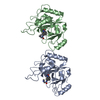
- Assembly
| Deposited unit | 
| ||||||||
|---|---|---|---|---|---|---|---|---|---|
| 1 | 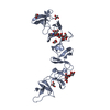
| ||||||||
| 2 | 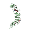
| ||||||||
| Unit cell |
|
-Components
| #1: Protein | Mass: 28644.740 Da / Num. of mol.: 2 / Fragment: TcdA fragment 2 Source method: isolated from a genetically manipulated source Source: (gene. exp.) Clostridium difficile (bacteria) / Strain: 48489 / Gene: toxA, tcdA / Plasmid: pET-3a / Production host: #2: Polysaccharide | alpha-D-galactopyranose-(1-3)-beta-D-galactopyranose-(1-4)-2-acetamido-2-deoxy-beta-D-glucopyranose Source method: isolated from a genetically manipulated source #3: Chemical | ChemComp-GOL /   Mass: 92.094 Da / Num. of mol.: 6 / Source method: obtained synthetically / Formula: C3H8O3 Mass: 92.094 Da / Num. of mol.: 6 / Source method: obtained synthetically / Formula: C3H8O3#4: Water | ChemComp-HOH / |  Mass: 18.015 Da / Num. of mol.: 413 / Source method: isolated from a natural source / Formula: H2O Mass: 18.015 Da / Num. of mol.: 413 / Source method: isolated from a natural source / Formula: H2O |
|---|
-Experimental details
-Experiment
| Experiment | Method: X-RAY DIFFRACTION / Number of used crystals: 1 |
|---|
- Sample preparation
| Crystal | Density Matthews: 2.91 Å3/Da / Density % sol: 57.7 % |
|---|---|
| Crystal grow | Temperature: 293 K / Method: vapor diffusion, hanging drop / pH: 7 Details: 6% PEG 3350, 0.1 M Bis-Tris-Cl pH 7.0, 5% glycerol, VAPOR DIFFUSION, HANGING DROP, temperature 293K |
-Data collection
| Diffraction | Mean temperature: 100 K |
|---|---|
| Diffraction source | Source: SYNCHROTRON / Site: ALS  / Beamline: 8.3.1 / Wavelength: 1.115 Å / Beamline: 8.3.1 / Wavelength: 1.115 Å |
| Detector | Type: ADSC QUANTUM 315 / Detector: CCD / Date: Aug 25, 2005 / Details: mirrors |
| Radiation | Monochromator: double crystal / Protocol: SINGLE WAVELENGTH / Monochromatic (M) / Laue (L): M / Scattering type: x-ray |
| Radiation wavelength | Wavelength: 1.115 Å / Relative weight: 1 |
| Reflection | Resolution: 2→60 Å / Num. all: 44075 / Num. obs: 44075 / % possible obs: 99.9 % / Observed criterion σ(F): -3 / Observed criterion σ(I): -3 / Redundancy: 4 % / Rmerge(I) obs: 0.047 / Rsym value: 0.047 / Net I/σ(I): 27.6 |
| Reflection shell | Resolution: 2→2.07 Å / Redundancy: 4 % / Rmerge(I) obs: 0.203 / Mean I/σ(I) obs: 7 / Num. unique all: 4692 / Rsym value: 0.203 / % possible all: 99.9 |
- Processing
| Software |
| |||||||||||||||||||||||||
|---|---|---|---|---|---|---|---|---|---|---|---|---|---|---|---|---|---|---|---|---|---|---|---|---|---|---|
| Refinement | Method to determine structure: MOLECULAR REPLACEMENT Starting model: PDB Entry 2F6E Resolution: 2→60 Å / Isotropic thermal model: Isotropic / Cross valid method: THROUGHOUT / σ(F): 0 / σ(I): 0 / Stereochemistry target values: Engh & Huber
| |||||||||||||||||||||||||
| Displacement parameters |
| |||||||||||||||||||||||||
| Refinement step | Cycle: LAST / Resolution: 2→60 Å
| |||||||||||||||||||||||||
| Refine LS restraints |
|
