negative regulation of DNA strand resection involved in replication fork processing / cell wall / SUMOylation of transcription factors / Recruitment and ATM-mediated phosphorylation of repair and signaling proteins at DNA double strand breaks / mitotic intra-S DNA damage checkpoint signaling / phosphorelay signal transduction system / mitotic G1 DNA damage checkpoint signaling / peptidoglycan-based cell wall / DNA damage checkpoint signaling / nucleotide-excision repair ...negative regulation of DNA strand resection involved in replication fork processing / cell wall / SUMOylation of transcription factors / Recruitment and ATM-mediated phosphorylation of repair and signaling proteins at DNA double strand breaks / mitotic intra-S DNA damage checkpoint signaling / phosphorelay signal transduction system / mitotic G1 DNA damage checkpoint signaling / peptidoglycan-based cell wall / DNA damage checkpoint signaling / nucleotide-excision repair / enzyme activator activity / double-strand break repair / double-stranded DNA binding / histone binding / regulation of cell cycle / DNA repair / GTPase activity / regulation of DNA-templated transcription / chromatin / positive regulation of DNA-templated transcription / positive regulation of transcription by RNA polymerase II / ATP hydrolysis activity / DNA binding / nucleus / plasma membrane Similarity search - Function
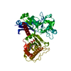
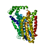
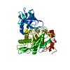
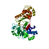
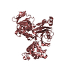
Mass: 41988.375 Da / Num. of mol.: 2 Source method: isolated from a genetically manipulated source Source: (gene. exp.) Mycobacterium tuberculosis (bacteria) / Strain: H37Rv / Gene: embR / Plasmid: pET23c / Production host: Escherichia coli (E. coli) / Strain (production host): C41(DE3) / References: UniProt: P66799, UniProt: P9WGJ9*PLUS
#2: Protein/peptide
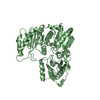
DNArepairproteinRAD9
Mass: 1043.963 Da / Num. of mol.: 2 / Source method: obtained synthetically Details: The sequence of the peptide is naturally found in Saccharomyces cerevisiae (yeast). References: UniProt: P14737
In the structure databanks used in Yorodumi, some data are registered as the other names, "COVID-19 virus" and "2019-nCoV". Here are the details of the virus and the list of structure data.
Jan 31, 2019. EMDB accession codes are about to change! (news from PDBe EMDB page)
EMDB accession codes are about to change! (news from PDBe EMDB page)
The allocation of 4 digits for EMDB accession codes will soon come to an end. Whilst these codes will remain in use, new EMDB accession codes will include an additional digit and will expand incrementally as the available range of codes is exhausted. The current 4-digit format prefixed with “EMD-” (i.e. EMD-XXXX) will advance to a 5-digit format (i.e. EMD-XXXXX), and so on. It is currently estimated that the 4-digit codes will be depleted around Spring 2019, at which point the 5-digit format will come into force.
The EM Navigator/Yorodumi systems omit the EMD- prefix.
Related info.:Q: What is EMD? / ID/Accession-code notation in Yorodumi/EM Navigator
Yorodumi is a browser for structure data from EMDB, PDB, SASBDB, etc.
This page is also the successor to EM Navigator detail page, and also detail information page/front-end page for Omokage search.
The word "yorodu" (or yorozu) is an old Japanese word meaning "ten thousand". "mi" (miru) is to see.
Related info.:EMDB / PDB / SASBDB / Comparison of 3 databanks / Yorodumi Search / Aug 31, 2016. New EM Navigator & Yorodumi / Yorodumi Papers / Jmol/JSmol / Function and homology information / Changes in new EM Navigator and Yorodumi
 Movie
Movie Controller

 Yorodumi
 Open data
 Basic information
 Components
 Keywords
 Function and homology information
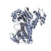
 Mycobacterium tuberculosis (bacteria)
 X-RAY DIFFRACTION /
  Authors
 Citation
 Structure visualization
 Downloads & links
 Other downloads
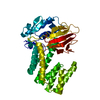

 PDBj
PDBj


 Assembly
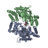
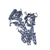

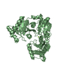
 Mass: 18.015 Da / Num. of mol.: 532 / Source method: isolated from a natural source / Formula: H2O
 Sample preparation
 / Beamline: ID29 / Wavelength: 0.9794 Å
 Processing