 Movie
Movie Controller

[English] 日本語
 Yorodumi
- PDB-2fdg: Crystal Structure of AlkB in complex with Fe(II), succinate, and ... -
+ Open data

- Basic information
| Entry | Database: PDB / ID: 2fdg | ||||||
|---|---|---|---|---|---|---|---|
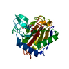
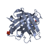
| Title | Crystal Structure of AlkB in complex with Fe(II), succinate, and methylated trinucleotide T-meA-T | ||||||
 Components Components |
| ||||||
 Keywords Keywords | OXIDOREDUCTASE/DNA / beta jellyroll / Structural Genomics / PSI / Protein Structure Initiative / Northeast Structural Genomics Consortium / NESG / OXIDOREDUCTASE-DNA COMPLEX | ||||||
| Function / homology |  Function and homology information Function and homology informationresponse to methyl methanesulfonate / oxidative RNA demethylation / DNA oxidative demethylase / broad specificity oxidative DNA demethylase activity / oxidative RNA demethylase activity / RNA repair / oxidative demethylation / DNA alkylation repair / dioxygenase activity / ferrous iron binding ...response to methyl methanesulfonate / oxidative RNA demethylation / DNA oxidative demethylase / broad specificity oxidative DNA demethylase activity / oxidative RNA demethylase activity / RNA repair / oxidative demethylation / DNA alkylation repair / dioxygenase activity / ferrous iron binding / DNA repair / cytoplasm / cytosol Similarity search - Function | ||||||
| Biological species |  | ||||||
| Method |  X-RAY DIFFRACTION / MOLECULAR REPLACEMENT / Resolution: 2.2 Å X-RAY DIFFRACTION / MOLECULAR REPLACEMENT / Resolution: 2.2 Å | ||||||
 Authors Authors | Yu, B. / Benach, J. / Edstrom, W.C. / Gibney, B.R. / Hunt, J.F. / Northeast Structural Genomics Consortium (NESG) | ||||||
 Citation Citation | Journal: Nature / Year: 2006 Title: Crystal structures of catalytic complexes of the oxidative DNA/RNA repair enzyme AlkB. Authors: Yu, B. / Edstrom, W.C. / Benach, J. / Hamuro, Y. / Weber, P.C. / Gibney, B.R. / Hunt, J.F. | ||||||
| History |
| ||||||
| Remark 600 | HETEROGEN The close contacts in remark 500 are due to iron coordination. |
- Structure visualization
| Structure viewer | Molecule: MolmilJmol/JSmol |
|---|
- Downloads & links
-Download
| PDBx/mmCIF format | 2fdg.cif.gz | 60.1 KB | Display | PDBx/mmCIF format |
|---|---|---|---|---|
| PDB format | pdb2fdg.ent.gz | 40.8 KB | Display | PDB format |
| PDBx/mmJSON format | 2fdg.json.gz | Tree view | PDBx/mmJSON format | |
| Others |  Other downloads Other downloads |
-Validation report
| Summary document | 2fdg_validation.pdf.gz | 450.7 KB | Display | wwPDB validaton report |
|---|---|---|---|---|
| Full document | 2fdg_full_validation.pdf.gz | 451.3 KB | Display | |
| Data in XML | 2fdg_validation.xml.gz | 11.2 KB | Display | |
| Data in CIF | 2fdg_validation.cif.gz | 15.3 KB | Display | |
| Arichive directory | https://data.pdbj.org/pub/pdb/validation_reports/fd/2fdgftp://data.pdbj.org/pub/pdb/validation_reports/fd/2fdg | HTTPS FTP |
-Related structure data
| Related structure data |  2fd8SC 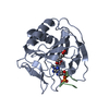 2fdfC  2fdhC  2fdiC 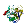 2fdjC  2fdkC S: Starting model for refinement C: citing same article ( |
|---|---|
| Similar structure data | |
| Other databases |


-Links
 PDBj
PDBj


- Assembly
| Deposited unit | 
| ||||||||
|---|---|---|---|---|---|---|---|---|---|
| 1 |
| ||||||||
| Unit cell |
|
-Components
| #1: DNA chain | Mass: 891.669 Da / Num. of mol.: 1 / Source method: obtained synthetically |
|---|---|
| #2: Protein | Mass: 23610.975 Da / Num. of mol.: 1 / Fragment: residues 12-216 Source method: isolated from a genetically manipulated source Source: (gene. exp.) |
| #3: Chemical | ChemComp-FE2 /   Mass: 55.845 Da / Num. of mol.: 1 / Source method: obtained synthetically / Formula: Fe Mass: 55.845 Da / Num. of mol.: 1 / Source method: obtained synthetically / Formula: Fe |
| #4: Chemical | ChemComp-SIN /   Mass: 118.088 Da / Num. of mol.: 1 / Source method: obtained synthetically / Formula: C4H6O4 Mass: 118.088 Da / Num. of mol.: 1 / Source method: obtained synthetically / Formula: C4H6O4 |
| #5: Water | ChemComp-HOH /  Mass: 18.015 Da / Num. of mol.: 119 / Source method: isolated from a natural source / Formula: H2O Mass: 18.015 Da / Num. of mol.: 119 / Source method: isolated from a natural source / Formula: H2O |
-Experimental details
-Experiment
| Experiment | Method: X-RAY DIFFRACTION / Number of used crystals: 1 |
|---|
- Sample preparation
| Crystal | Density Matthews: 1.96 Å3/Da / Density % sol: 37.23 % |
|---|---|
| Crystal grow | Temperature: 298 K / Method: vapor diffusion, hanging drop Details: 22-24% (w/v) PEG 3350, 10% glycerol and 200 mM sodium formate, VAPOR DIFFUSION, HANGING DROP, temperature 298K |
-Data collection
| Diffraction | Mean temperature: 100 K |
|---|---|
| Diffraction source | Source: ROTATING ANODE / Type: RIGAKU RU200 / Wavelength: 1.5418 Å |
| Detector | Type: RIGAKU RAXIS IV / Detector: IMAGE PLATE / Date: Aug 17, 2005 / Details: Yale/MSC mirrors |
| Radiation | Protocol: SINGLE WAVELENGTH / Monochromatic (M) / Laue (L): M / Scattering type: x-ray |
| Radiation wavelength | Wavelength: 1.5418 Å / Relative weight: 1 |
| Reflection | Resolution: 2.2→20 Å / Num. obs: 9504 / % possible obs: 99 % / Redundancy: 4.8 % / Rmerge(I) obs: 0.089 / Χ2: 1.026 / Net I/σ(I): 17.77 |
| Reflection shell | Resolution: 2.2→2.28 Å / Redundancy: 3.9 % / Rmerge(I) obs: 0.304 / Mean I/σ(I) obs: 4.8 / Num. unique all: 932 / Χ2: 1.258 / % possible all: 98.3 |
- Processing
| Software |
| ||||||||||||||||||||||||||||||||||||
|---|---|---|---|---|---|---|---|---|---|---|---|---|---|---|---|---|---|---|---|---|---|---|---|---|---|---|---|---|---|---|---|---|---|---|---|---|---|
| Refinement | Method to determine structure: MOLECULAR REPLACEMENT Starting model: PDB 2FD8 Resolution: 2.2→19.94 Å / Rfactor Rfree error: 0.007 / Data cutoff high absF: 1079489.75 / Data cutoff low absF: 0 / Isotropic thermal model: RESTRAINED / Cross valid method: THROUGHOUT / σ(F): 1
| ||||||||||||||||||||||||||||||||||||
| Solvent computation | Solvent model: FLAT MODEL / Bsol: 41.185 Å2 / ksol: 0.367 e/Å3 | ||||||||||||||||||||||||||||||||||||
| Displacement parameters | Biso mean: 19.1 Å2
| ||||||||||||||||||||||||||||||||||||
| Refine analyze |
| ||||||||||||||||||||||||||||||||||||
| Refinement step | Cycle: LAST / Resolution: 2.2→19.94 Å
| ||||||||||||||||||||||||||||||||||||
| Refine LS restraints |
| ||||||||||||||||||||||||||||||||||||
| LS refinement shell | Resolution: 2.2→2.34 Å / Rfactor Rfree error: 0.022 / Total num. of bins used: 6
| ||||||||||||||||||||||||||||||||||||
| Xplor file |
|
