 Movie
Movie Controller

[English] 日本語
 Yorodumi
- PDB-2f3o: Crystal Structure of a glycyl radical enzyme from Archaeoglobus f... -
+ Open data

- Basic information
| Entry | Database: PDB / ID: 2f3o | ||||||
|---|---|---|---|---|---|---|---|
| Title | Crystal Structure of a glycyl radical enzyme from Archaeoglobus fulgidus | ||||||
 Components Components | pyruvate formate-lyase 2 | ||||||
 Keywords Keywords | UNKNOWN FUNCTION / pyruvate formate lyase / glycerol dehydratase / PFL2 / glycyl radical / hyperthermophilic | ||||||
| Function / homology |  Function and homology information Function and homology information | ||||||
| Biological species |   Archaeoglobus fulgidus (archaea) Archaeoglobus fulgidus (archaea) | ||||||
| Method |  X-RAY DIFFRACTION / SYNCHROTRON / MOLECULAR REPLACEMENT / Resolution: 2.9 Å X-RAY DIFFRACTION / SYNCHROTRON / MOLECULAR REPLACEMENT / Resolution: 2.9 Å | ||||||
 Authors Authors | Lehtio, L. / Goldman, A. | ||||||
 Citation Citation | Journal: J.Mol.Biol. / Year: 2006 Title: Crystal structure of a glycyl radical enzyme from Archaeoglobus fulgidus Authors: Lehtio, L. / Grossmann, J.G. / Kokona, B. / Fairman, R. / Goldman, A. | ||||||
| History |
|
- Structure visualization
| Structure viewer | Molecule: MolmilJmol/JSmol |
|---|
- Downloads & links
-Download
| PDBx/mmCIF format | 2f3o.cif.gz | 307.9 KB | Display | PDBx/mmCIF format |
|---|---|---|---|---|
| PDB format | pdb2f3o.ent.gz | 251.1 KB | Display | PDB format |
| PDBx/mmJSON format | 2f3o.json.gz | Tree view | PDBx/mmJSON format | |
| Others |  Other downloads Other downloads |
-Validation report
| Arichive directory | https://data.pdbj.org/pub/pdb/validation_reports/f3/2f3oftp://data.pdbj.org/pub/pdb/validation_reports/f3/2f3o | HTTPS FTP |
|---|
-Related structure data
| Related structure data |  1r9dS S: Starting model for refinement |
|---|---|
| Similar structure data |

-Links
 PDBj
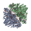
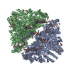
PDBj- Assembly
| Deposited unit | 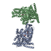
| |||||||||||||||
|---|---|---|---|---|---|---|---|---|---|---|---|---|---|---|---|---|
| 1 | 
| |||||||||||||||
| 2 | 
| |||||||||||||||
| Unit cell |
| |||||||||||||||
| Components on special symmetry positions |
| |||||||||||||||
| Details | The biological tetramer is formed from chain A with symmetry operators: -x, -y+1, z x, -y+1, -z+1 -x, y, -z+1 / The biological tetramer is formed from chain B with symmetry operators: x, -y+1, -z -x-1, -y+1, z -x-1, y, -z+2 |
-Components
| #1: Protein | Mass: 87264.477 Da / Num. of mol.: 2 Source method: isolated from a genetically manipulated source Source: (gene. exp.) Archaeoglobus fulgidus (archaea) / Gene: AF1449, PFLD / Plasmid: pQE-30 / Production host:  #2: Chemical |   Mass: 150.173 Da / Num. of mol.: 2 / Source method: obtained synthetically / Formula: C6H14O4 Mass: 150.173 Da / Num. of mol.: 2 / Source method: obtained synthetically / Formula: C6H14O4#3: Chemical |   Mass: 92.094 Da / Num. of mol.: 2 / Source method: obtained synthetically / Formula: C3H8O3 Mass: 92.094 Da / Num. of mol.: 2 / Source method: obtained synthetically / Formula: C3H8O3#4: Water | ChemComp-HOH / |  Mass: 18.015 Da / Num. of mol.: 167 / Source method: isolated from a natural source / Formula: H2O Mass: 18.015 Da / Num. of mol.: 167 / Source method: isolated from a natural source / Formula: H2O |
|---|
-Experimental details
-Experiment
| Experiment | Method: X-RAY DIFFRACTION / Number of used crystals: 1 |
|---|
- Sample preparation
| Crystal | Density Matthews: 3.38 Å3/Da / Density % sol: 63.66 % |
|---|---|
| Crystal grow | Temperature: 298 K / Method: vapor diffusion, sitting drop / pH: 7.5 Details: 16 % PEG8000, 8 % isopropanol, 80 mM Hepes pH 7.5, 160 mM ammonium sulphate, 1 mM DTT, VAPOR DIFFUSION, SITTING DROP, temperature 298.0K |
-Data collection
| Diffraction | Mean temperature: 100 K |
|---|---|
| Diffraction source | Source: SYNCHROTRON / Site: ESRF  / Beamline: ID29 / Wavelength: 1 Å / Beamline: ID29 / Wavelength: 1 Å |
| Detector | Type: ADSC QUANTUM 4 / Detector: CCD / Date: Feb 21, 2004 |
| Radiation | Monochromator: Si 111 CHANNEL / Protocol: SINGLE WAVELENGTH / Monochromatic (M) / Laue (L): M / Scattering type: x-ray |
| Radiation wavelength | Wavelength: 1 Å / Relative weight: 1 |
| Reflection | Resolution: 2.9→20 Å / Num. all: 52531 / Num. obs: 52531 / % possible obs: 98.6 % / Observed criterion σ(F): 0 / Observed criterion σ(I): -3 / Rmerge(I) obs: 0.087 / Net I/σ(I): 20.61 |
| Reflection shell | Resolution: 2.9→3 Å / Rmerge(I) obs: 0.449 / Mean I/σ(I) obs: 5.34 / Num. unique all: 36376 / Num. unique obs: 4896 / % possible all: 97.2 |
-Phasing
| Phasing MR | Cor.coef. Fo:Fc: 0.185 / Packing: 0.427
|
|---|
- Processing
| Software |
| ||||||||||||||||||||||||||||||||||||||||||||||||||||||||||||||||||||||||||||||||||||||||||
|---|---|---|---|---|---|---|---|---|---|---|---|---|---|---|---|---|---|---|---|---|---|---|---|---|---|---|---|---|---|---|---|---|---|---|---|---|---|---|---|---|---|---|---|---|---|---|---|---|---|---|---|---|---|---|---|---|---|---|---|---|---|---|---|---|---|---|---|---|---|---|---|---|---|---|---|---|---|---|---|---|---|---|---|---|---|---|---|---|---|---|---|
| Refinement | Method to determine structure: MOLECULAR REPLACEMENT Starting model: pdb entry 1R9D Resolution: 2.9→19.69 Å / Cor.coef. Fo:Fc: 0.938 / Cor.coef. Fo:Fc free: 0.909 / SU B: 16.048 / SU ML: 0.288 / Cross valid method: THROUGHOUT / σ(F): 0 / ESU R: 4.668 / ESU R Free: 0.361 / Stereochemistry target values: MAXIMUM LIKELIHOOD
| ||||||||||||||||||||||||||||||||||||||||||||||||||||||||||||||||||||||||||||||||||||||||||
| Solvent computation | Ion probe radii: 0.8 Å / Shrinkage radii: 0.8 Å / VDW probe radii: 1.2 Å / Solvent model: MASK | ||||||||||||||||||||||||||||||||||||||||||||||||||||||||||||||||||||||||||||||||||||||||||
| Displacement parameters | Biso mean: 57.512 Å2
| ||||||||||||||||||||||||||||||||||||||||||||||||||||||||||||||||||||||||||||||||||||||||||
| Refinement step | Cycle: LAST / Resolution: 2.9→19.69 Å
| ||||||||||||||||||||||||||||||||||||||||||||||||||||||||||||||||||||||||||||||||||||||||||
| Refine LS restraints |
| ||||||||||||||||||||||||||||||||||||||||||||||||||||||||||||||||||||||||||||||||||||||||||
| LS refinement shell | Resolution: 2.9→2.974 Å / Total num. of bins used: 20
|
