 Movie
Movie Controller

[English] 日本語
 Yorodumi
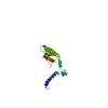
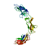
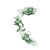
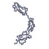
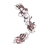
- PDB-2f3j: The solution structure of the REF2-I mRNA export factor (residues... -
+ Open data

- Basic information
| Entry | Database: PDB / ID: 2f3j | ||||||
|---|---|---|---|---|---|---|---|
| Title | The solution structure of the REF2-I mRNA export factor (residues 1-155). | ||||||
 Components Components | RNA and export factor binding protein 2 | ||||||
 Keywords Keywords | TRANSPORT PROTEIN / RRM domain / RBD domain. | ||||||
| Function / homology |  Function and homology information Function and homology informationmRNA transport / RNA splicing / spliceosomal complex / mRNA processing / single-stranded DNA binding / RNA binding / cytoplasm Similarity search - Function | ||||||
| Biological species |  | ||||||
| Method | SOLUTION NMR / ARIA protocol (Nilges et al., 1997, J. Mol. Biol. 269, 408-422) used for structure calculation. | ||||||
 Authors Authors | Golovanov, A.P. / Hautbergue, G.M. / Wilson, S.A. / Lian, L.Y. | ||||||
 Citation Citation | Journal: Rna / Year: 2006 Title: The solution structure of REF2-I reveals interdomain interactions and regions involved in binding mRNA export factors and RNA. Authors: Golovanov, A.P. / Hautbergue, G.M. / Tintaru, A.M. / Lian, L.Y. / Wilson, S.A. | ||||||
| History |
|
- Structure visualization
| Structure viewer | Molecule: MolmilJmol/JSmol |
|---|
- Downloads & links
-Download
| PDBx/mmCIF format | 2f3j.cif.gz | 758 KB | Display | PDBx/mmCIF format |
|---|---|---|---|---|
| PDB format | pdb2f3j.ent.gz | 642.6 KB | Display | PDB format |
| PDBx/mmJSON format | 2f3j.json.gz | Tree view | PDBx/mmJSON format | |
| Others |  Other downloads Other downloads |
-Validation report
| Arichive directory | https://data.pdbj.org/pub/pdb/validation_reports/f3/2f3jftp://data.pdbj.org/pub/pdb/validation_reports/f3/2f3j | HTTPS FTP |
|---|
-Related structure data
| Similar structure data |
|---|


-Links
 PDBj
PDBj- Assembly
| Deposited unit | 
| |||||||||
|---|---|---|---|---|---|---|---|---|---|---|
| 1 |
| |||||||||
| NMR ensembles |
|
-Components
| #1: Protein | Mass: 19630.188 Da / Num. of mol.: 1 / Fragment: RRM Domain (Residues 1 - 155) Source method: isolated from a genetically manipulated source Source: (gene. exp.)  |
|---|
-Experimental details
-Experiment
| Experiment | Method: SOLUTION NMR | ||||||||||||||||||||
|---|---|---|---|---|---|---|---|---|---|---|---|---|---|---|---|---|---|---|---|---|---|
| NMR experiment |
| ||||||||||||||||||||
| NMR details | Text: The protein construct contains 14-residue N-terminal T7 tag (MASMTGGQQMGRDP), and C-terminal tag (LEHHHHHH). The T7 tag is unstructured and is omitted in the coordinate file, where residue ...Text: The protein construct contains 14-residue N-terminal T7 tag (MASMTGGQQMGRDP), and C-terminal tag (LEHHHHHH). The T7 tag is unstructured and is omitted in the coordinate file, where residue numbering starts from residue 1 of REF2-I. Last three residues (LEH) in the coordinate file originate from the beginning of C-terminal tag. The N-terminal domain (1-74) of REF2-I is flexible and is largely unstructured, apart from the region 8-18 which forms a transient helix. |
 HSQC
HSQC- Sample preparation
| Details |
| |||||||||||||||
|---|---|---|---|---|---|---|---|---|---|---|---|---|---|---|---|---|
| Sample conditions |
|
-NMR measurement
| Radiation | Protocol: SINGLE WAVELENGTH / Monochromatic (M) / Laue (L): M | |||||||||||||||
|---|---|---|---|---|---|---|---|---|---|---|---|---|---|---|---|---|
| Radiation wavelength | Relative weight: 1 | |||||||||||||||
| NMR spectrometer |
|
- Processing
| NMR software |
| ||||||||||||||||||||||||||||
|---|---|---|---|---|---|---|---|---|---|---|---|---|---|---|---|---|---|---|---|---|---|---|---|---|---|---|---|---|---|
| Refinement | Method: ARIA protocol (Nilges et al., 1997, J. Mol. Biol. 269, 408-422) used for structure calculation. Software ordinal: 1 Details: Residual dipolar couplings were measured in 5% PEG liquid crystal media (C12E5 + hexane, Ruckert and Otting, 2000, J. Am. Chem. Soc. 122, 7793) and used as SANI restraints in ARIA. | ||||||||||||||||||||||||||||
| NMR representative | Selection criteria: lowest energy | ||||||||||||||||||||||||||||
| NMR ensemble | Conformer selection criteria: structures with the lowest energy Conformers calculated total number: 20 / Conformers submitted total number: 14 |
