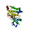
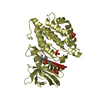
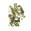
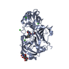
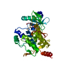
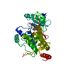
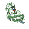
Entry Database : PDB / ID : 2evaTitle Structural Basis for the Interaction of TAK1 Kinase with its Activating Protein TAB1 TAK1 kinase - TAB1 chimera fusion protein Keywords / / / / Function / homology Function Domain/homology Component
/ / / / / / / / / / / / / / / / / / / / / / / / / / / / / / / / / / / / / / / / / / / / / / / / / / / / / / / / / / / / / / / / / / / / / / / / / / / / / / / / / / / / / / / / / / / / / / / / / / / / / / / / / / / / / / / / / / / / / / Biological species Homo sapiens (human)Method / / / Resolution : 2 Å Authors Brown, K. / Vial, S.C. / Dedi, N. / Long, J.M. / Dunster, N.J. / Cheetham, G.M. Journal : J.Mol.Biol. / Year : 2005Title : Structural basis for the interaction of TAK1 kinase with its activating protein TAB1Authors : Brown, K. / Vial, S.C. / Dedi, N. / Long, J.M. / Dunster, N.J. / Cheetham, G.M. History Deposition Oct 31, 2005 Deposition site / Processing site Revision 1.0 May 2, 2006 Provider / Type Revision 1.1 May 1, 2008 Group Revision 1.2 Jul 13, 2011 Group Revision 1.3 Aug 2, 2017 Group / Source and taxonomy / Category / softwareRevision 1.4 Oct 18, 2017 Group / Category / Item Revision 1.5 Feb 14, 2024 Group / Database references / Derived calculationsCategory chem_comp_atom / chem_comp_bond ... chem_comp_atom / chem_comp_bond / database_2 / struct_ref_seq_dif / struct_site Item _database_2.pdbx_DOI / _database_2.pdbx_database_accession ... _database_2.pdbx_DOI / _database_2.pdbx_database_accession / _struct_ref_seq_dif.details / _struct_site.pdbx_auth_asym_id / _struct_site.pdbx_auth_comp_id / _struct_site.pdbx_auth_seq_id
Show all Show less
 Movie
Movie Controller

 Yorodumi
 Open data
 Basic information
 Components
 Keywords
 Function and homology information
 Homo sapiens (human)
 X-RAY DIFFRACTION /
  Authors
 Citation
 Structure visualization
 Downloads & links
 Other downloads
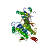


 PDBj
PDBj


 Assembly

 Spodoptera frugiperda (fall armyworm) / References: UniProt: O43318, EC: 2.7.1.37
 Mass: 267.241 Da / Num. of mol.: 1 / Source method: obtained synthetically / Formula: C10H13N5O4
 Mass: 18.015 Da / Num. of mol.: 229 / Source method: isolated from a natural source / Formula: H2O
 Sample preparation
 / Beamline: PX14.1 / Wavelength: 1.488 Å
 Processing