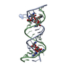
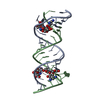
 Movie
Movie Controller


+ Open data

- Basic information
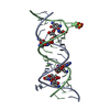
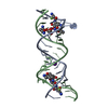
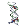
| Entry | Database: PDB / ID: 2et3 | ||||||||||||||||||
|---|---|---|---|---|---|---|---|---|---|---|---|---|---|---|---|---|---|---|---|
| Title | Complex Between Gentamicin C1A and the 16S-RRNA A-Site | ||||||||||||||||||
 Components Components | 5'-R(* Keywords KeywordsRNA / RNA-AMINOGLYCOSIDE INTERACTIONS / A SITE / UOU PAIRS / AA BULGES | Function / homology | Chem-LLL / RNA / RNA (> 10) |  Function and homology information Function and homology informationMethod |  X-RAY DIFFRACTION / SYNCHROTRON / MOLECULAR REPLACEMENT / Resolution: 2.8 Å X-RAY DIFFRACTION / SYNCHROTRON / MOLECULAR REPLACEMENT / Resolution: 2.8 Å  Authors AuthorsWesthof, E. |  CitationJournal: Nucleic Acids Res. / Year: 2005 CitationJournal: Nucleic Acids Res. / Year: 2005Title: Crystal structures of complexes between aminoglycosides and decoding A site oligonucleotides: role of the number of rings and positive charges in the specific binding leading to miscoding. Authors: Francois, B. / Russell, R.J. / Murray, J.B. / Aboul-ela, F. / Masquida, B. / Vicens, Q. / Westhof, E. History |
|
- Structure visualization
| Structure viewer | Molecule: MolmilJmol/JSmol |
|---|
- Downloads & links
-Download
| PDBx/mmCIF format | 2et3.cif.gz | 35.4 KB | Display | PDBx/mmCIF format |
|---|---|---|---|---|
| PDB format | pdb2et3.ent.gz | 24 KB | Display | PDB format |
| PDBx/mmJSON format | 2et3.json.gz | Tree view | PDBx/mmJSON format | |
| Others |  Other downloads Other downloads |
-Validation report
| Arichive directory | https://data.pdbj.org/pub/pdb/validation_reports/et/2et3ftp://data.pdbj.org/pub/pdb/validation_reports/et/2et3 | HTTPS FTP |
|---|
-Related structure data
| Related structure data |  2esiC  2esjC  2et4C  2et5C 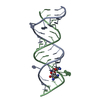 2et8C 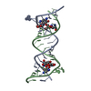 1j7tS S: Starting model for refinement C: citing same article ( |
|---|---|
| Similar structure data |


-Links
 PDBj
PDBj


- Assembly
| Deposited unit | 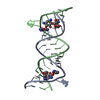
| ||||||||
|---|---|---|---|---|---|---|---|---|---|
| 1 |
| ||||||||
| Unit cell |
|
-Components
| #1: RNA chain | Mass: 7048.259 Da / Num. of mol.: 2 / Source method: obtained synthetically #2: Chemical | 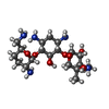  Mass: 449.542 Da / Num. of mol.: 2 / Source method: obtained synthetically / Formula: C19H39N5O7 Mass: 449.542 Da / Num. of mol.: 2 / Source method: obtained synthetically / Formula: C19H39N5O7#3: Water | ChemComp-HOH / |  Mass: 18.015 Da / Num. of mol.: 8 / Source method: isolated from a natural source / Formula: H2O Mass: 18.015 Da / Num. of mol.: 8 / Source method: isolated from a natural source / Formula: H2O |
|---|
-Experimental details
-Experiment
| Experiment | Method: X-RAY DIFFRACTION / Number of used crystals: 1 |
|---|
- Sample preparation
| Crystal | Density Matthews: 2.51 Å3/Da / Density % sol: 50.99 % | ||||||||||||||||||||||||||||||||||||||||||||
|---|---|---|---|---|---|---|---|---|---|---|---|---|---|---|---|---|---|---|---|---|---|---|---|---|---|---|---|---|---|---|---|---|---|---|---|---|---|---|---|---|---|---|---|---|---|
| Crystal grow | Temperature: 310 K / Method: vapor diffusion, hanging drop / pH: 6.4 Details: MPD, NACL, MGSO4, GLYCEROL, NA CACODYLATE, pH 6.4, VAPOR DIFFUSION, HANGING DROP, temperature 310K | ||||||||||||||||||||||||||||||||||||||||||||
| Components of the solutions |
|
-Data collection
| Diffraction | Mean temperature: 110 K |
|---|---|
| Diffraction source | Source: SYNCHROTRON / Site: ESRF  / Beamline: ID29 / Wavelength: 0.936 Å / Beamline: ID29 / Wavelength: 0.936 Å |
| Detector | Type: ADSC QUANTUM 4 / Detector: CCD |
| Radiation | Protocol: SINGLE WAVELENGTH / Monochromatic (M) / Laue (L): M / Scattering type: x-ray |
| Radiation wavelength | Wavelength: 0.936 Å / Relative weight: 1 |
| Reflection | Resolution: 2.8→50 Å / Num. all: 4000 / Num. obs: 4000 / Observed criterion σ(F): 5 / Observed criterion σ(I): 5 |
- Processing
| Software |
| |||||||||||||||||||||||||
|---|---|---|---|---|---|---|---|---|---|---|---|---|---|---|---|---|---|---|---|---|---|---|---|---|---|---|
| Refinement | Method to determine structure: MOLECULAR REPLACEMENT Starting model: PDB Entry: 1J7T Resolution: 2.8→20 Å / σ(F): 1.5 Stereochemistry target values: G. PARKINSON, J. VOJTECHOVSKY, L. CLOWNEY,A.T. BRUNGER, H.M. BERMAN, NEW PARAMETERSFOR THE REFINEMENT OF NUCLEIC ACID CONTAINING STRUCTURES, ACTA CRYST. D, 52,57-64 (1996)
| |||||||||||||||||||||||||
| Refine analyze |
| |||||||||||||||||||||||||
| Refinement step | Cycle: LAST / Resolution: 2.8→20 Å
| |||||||||||||||||||||||||
| Refine LS restraints |
|
