Resolution: 1.4→35.71 Å / Cor.coef. Fo:Fc: 0.961 / Cor.coef. Fo:Fc free: 0.962 / SU B: 1.712 / SU ML: 0.034 / TLS residual ADP flag: LIKELY RESIDUAL / Isotropic thermal model: Isotropic / Cross valid method: THROUGHOUT / σ(F): 0 / ESU R: 0.055 / ESU R Free: 0.053 / Stereochemistry target values: MAXIMUM LIKELIHOOD Details: SIMULATED ANNEALING WAS PERFORMED WITH THE CNS SOFTWARE IN THE FIRST CYCLE OF REFINEMENT. TLS APPLIED USING THE PROTEIN MODEL AS ONE GROUP. Close contacts in remark 500 are related to the ...Details: SIMULATED ANNEALING WAS PERFORMED WITH THE CNS SOFTWARE IN THE FIRST CYCLE OF REFINEMENT. TLS APPLIED USING THE PROTEIN MODEL AS ONE GROUP. Close contacts in remark 500 are related to the alternate conformations of the CYS 6 region close to the N-terminus and CYS 31. The alternate conformation of the N-terminus is not modelled due to the weak electron density.
Rfactor
Num. reflection
% reflection
Selection details
Rfree
0.1895
676
5 %
RANDOM
Rwork
0.18014
-
-
-
all
0.1806
13610
-
-
obs
0.1806
13574
99.75 %
-
Solvent computation
Ion probe radii: 0.8 Å / Shrinkage radii: 0.8 Å / VDW probe radii: 1.2 Å / Solvent model: BABINET MODEL WITH MASK
Displacement parameters
Biso mean: 24.051 Å2
Baniso -1
Baniso -2
Baniso -3
1-
0.03 Å2
0 Å2
0 Å2
2-
-
0.21 Å2
0 Å2
3-
-
-
-0.24 Å2
Refinement step
Cycle: LAST / Resolution: 1.4→35.71 Å
Protein
Nucleic acid
Ligand
Solvent
Total
Num. atoms
402
0
0
74
476
Refine LS restraints
Refine-ID
Type
Dev ideal
Dev ideal target
Number
X-RAY DIFFRACTION
r_bond_refined_d
0.014
0.022
432
X-RAY DIFFRACTION
r_angle_refined_deg
1.486
1.968
582
X-RAY DIFFRACTION
r_dihedral_angle_1_deg
5.936
5
52
X-RAY DIFFRACTION
r_dihedral_angle_2_deg
35.224
23.81
21
X-RAY DIFFRACTION
r_dihedral_angle_3_deg
14.454
15
79
X-RAY DIFFRACTION
r_dihedral_angle_4_deg
15.321
15
4
X-RAY DIFFRACTION
r_chiral_restr
0.083
0.2
59
X-RAY DIFFRACTION
r_gen_planes_refined
0.008
0.02
332
X-RAY DIFFRACTION
r_nbd_refined
0.244
0.2
212
X-RAY DIFFRACTION
r_nbtor_refined
0.322
0.2
308
X-RAY DIFFRACTION
r_xyhbond_nbd_refined
0.127
0.2
91
X-RAY DIFFRACTION
r_symmetry_vdw_refined
0.177
0.2
44
X-RAY DIFFRACTION
r_symmetry_hbond_refined
0.146
0.2
8
X-RAY DIFFRACTION
r_mcbond_it
0.74
1.5
269
X-RAY DIFFRACTION
r_mcangle_it
1.219
2
430
X-RAY DIFFRACTION
r_scbond_it
1.764
3
178
X-RAY DIFFRACTION
r_scangle_it
2.954
4.5
152
LS refinement shell
Resolution: 1.403→1.439 Å / Total num. of bins used: 20
Rfactor
Num. reflection
% reflection
Rfree
0.38
55
-
Rwork
0.329
904
-
obs
-
904
97.96 %
Refinement TLS params.
Method: refined / Origin x: 12.833 Å / Origin y: 7.551 Å / Origin z: 24.913 Å
11
12
13
21
22
23
31
32
33
T
-0.1661 Å2
0.0011 Å2
0.0157 Å2
-
-0.1594 Å2
-0.0078 Å2
-
-
-0.1346 Å2
L
3.33 °2
0.357 °2
0.3214 °2
-
3.8251 °2
0.189 °2
-
-
1.6965 °2
S
-0.0121 Å °
0.0534 Å °
-0.1421 Å °
-0.1988 Å °
0.0332 Å °
-0.1068 Å °
0.1021 Å °
0.0824 Å °
-0.0211 Å °
+
About Yorodumi
-
News
-
Feb 9, 2022. New format data for meta-information of EMDB entries
New format data for meta-information of EMDB entries
Version 3 of the EMDB header file is now the official format.
The previous official version 1.9 will be removed from the archive.
In the structure databanks used in Yorodumi, some data are registered as the other names, "COVID-19 virus" and "2019-nCoV". Here are the details of the virus and the list of structure data.
Jan 31, 2019. EMDB accession codes are about to change! (news from PDBe EMDB page)
EMDB accession codes are about to change! (news from PDBe EMDB page)
The allocation of 4 digits for EMDB accession codes will soon come to an end. Whilst these codes will remain in use, new EMDB accession codes will include an additional digit and will expand incrementally as the available range of codes is exhausted. The current 4-digit format prefixed with “EMD-” (i.e. EMD-XXXX) will advance to a 5-digit format (i.e. EMD-XXXXX), and so on. It is currently estimated that the 4-digit codes will be depleted around Spring 2019, at which point the 5-digit format will come into force.
The EM Navigator/Yorodumi systems omit the EMD- prefix.
Related info.:Q: What is EMD? / ID/Accession-code notation in Yorodumi/EM Navigator
Yorodumi is a browser for structure data from EMDB, PDB, SASBDB, etc.
This page is also the successor to EM Navigator detail page, and also detail information page/front-end page for Omokage search.
The word "yorodu" (or yorozu) is an old Japanese word meaning "ten thousand". "mi" (miru) is to see.
Related info.:EMDB / PDB / SASBDB / Comparison of 3 databanks / Yorodumi Search / Aug 31, 2016. New EM Navigator & Yorodumi / Yorodumi Papers / Jmol/JSmol / Function and homology information / Changes in new EM Navigator and Yorodumi
 Movie
Movie Controller


 Open data
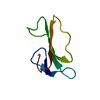
 Basic information
 Components
 Keywords
 Function and homology information
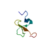
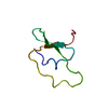
 Triatoma infestans (insect)
 X-RAY DIFFRACTION /
  Authors
 Citation
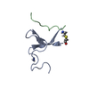
 Structure visualization
 Downloads & links
 Other downloads


 PDBj
PDBj
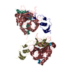
 Assembly
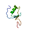
 Pichia pastoris (fungus) / Strain (production host): GS115 / References: UniProt: Q95P16
 Mass: 18.015 Da / Num. of mol.: 74 / Source method: isolated from a natural source / Formula: H2O
 Sample preparation
 / Beamline: D03B-MX1 / Wavelength: 1.427 Å
 Processing