 Movie
Movie Controller

[English] 日本語
 Yorodumi
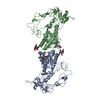
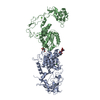
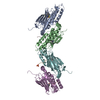
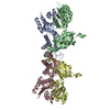
- PDB-2ero: Crystal structure of vascular apoptosis-inducing protein-1(orthor... -
+ Open data

- Basic information
| Entry | Database: PDB / ID: 2ero | |||||||||
|---|---|---|---|---|---|---|---|---|---|---|
| Title | Crystal structure of vascular apoptosis-inducing protein-1(orthorhombic crystal form) | |||||||||
 Components Components | vascular apoptosis-inducing protein 1 | |||||||||
 Keywords Keywords | TOXIN / metalloprotease / disintegrin / calcium-binding / ADAM / SVMP / MDC protein | |||||||||
| Function / homology |  Function and homology information Function and homology informationHydrolases; Acting on peptide bonds (peptidases); Metalloendopeptidases / metalloendopeptidase activity / toxin activity / apoptotic process / protein homodimerization activity / proteolysis / extracellular region / zinc ion binding / identical protein binding / plasma membrane Similarity search - Function | |||||||||
| Biological species |  Crotalus atrox (western diamondback rattlesnake) Crotalus atrox (western diamondback rattlesnake) | |||||||||
| Method |  X-RAY DIFFRACTION / SYNCHROTRON / MOLECULAR REPLACEMENT / Resolution: 2.5 Å X-RAY DIFFRACTION / SYNCHROTRON / MOLECULAR REPLACEMENT / Resolution: 2.5 Å | |||||||||
 Authors Authors | Takeda, S. / Igarashi, T. / Araki, S. | |||||||||
 Citation Citation | Journal: Embo J. / Year: 2006 Title: Crystal structures of VAP1 reveal ADAMs' MDC domain architecture and its unique C-shaped scaffold Authors: Takeda, S. / Igarashi, T. / Mori, H. / Araki, S. #1: Journal: To be PublishedTitle: Crystallization and preliminary X-ray crystallographic analysis of two vascular apoptosis-inducing proteins (VAPs) from Crotalus atrox venom Authors: Igarashi, T. / Oishi, Y. / Araki, S. / Mori, H. / Takeda, S. | |||||||||
| History |
|
- Structure visualization
| Structure viewer | Molecule: MolmilJmol/JSmol |
|---|
- Downloads & links
-Download
| PDBx/mmCIF format | 2ero.cif.gz | 183 KB | Display | PDBx/mmCIF format |
|---|---|---|---|---|
| PDB format | pdb2ero.ent.gz | 143.9 KB | Display | PDB format |
| PDBx/mmJSON format | 2ero.json.gz | Tree view | PDBx/mmJSON format | |
| Others |  Other downloads Other downloads |
-Validation report
| Arichive directory | https://data.pdbj.org/pub/pdb/validation_reports/er/2eroftp://data.pdbj.org/pub/pdb/validation_reports/er/2ero | HTTPS FTP |
|---|
-Related structure data
| Related structure data |  2erpC  2erqC  1quaS C: citing same article ( S: Starting model for refinement |
|---|---|
| Similar structure data |
-Links
 PDBj
PDBj


- Assembly
| Deposited unit | 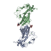
| ||||||||
|---|---|---|---|---|---|---|---|---|---|
| 1 |
| ||||||||
| 2 | 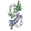
| ||||||||
| Unit cell |
| ||||||||
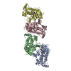
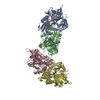
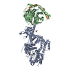
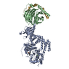
| Details | Asymmetric unit contains one disulfide-linked homodimer molecule that is the biological unit. |
-Components
-Protein / Sugars , 2 types, 4 molecules AB
| #1: Protein | Mass: 47378.000 Da / Num. of mol.: 2 / Fragment: residues 184-610 / Source method: isolated from a natural source Source: (natural) Crotalus atrox (western diamondback rattlesnake)Secretion: venom / References: GenBank: 11320556, UniProt: Q9DGB9*PLUS #2: Polysaccharide | Source method: isolated from a genetically manipulated source |
|---|
-Non-polymers , 4 types, 210 molecules 


| #3: Chemical |  Mass: 65.409 Da / Num. of mol.: 2 / Source method: obtained synthetically / Formula: Zn Mass: 65.409 Da / Num. of mol.: 2 / Source method: obtained synthetically / Formula: Zn#4: Chemical | ChemComp-CA /  Mass: 40.078 Da / Num. of mol.: 4 / Source method: obtained synthetically / Formula: Ca Mass: 40.078 Da / Num. of mol.: 4 / Source method: obtained synthetically / Formula: Ca#5: Chemical | ChemComp-3CO / |  Mass: 58.933 Da / Num. of mol.: 1 / Source method: obtained synthetically / Formula: Co Mass: 58.933 Da / Num. of mol.: 1 / Source method: obtained synthetically / Formula: Co#6: Water | ChemComp-HOH / | Mass: 18.015 Da / Num. of mol.: 203 / Source method: isolated from a natural source / Formula: H2O |
|---|
-Details
| Has protein modification | Y |
|---|
-Experimental details
-Experiment
| Experiment | Method: X-RAY DIFFRACTION / Number of used crystals: 1 |
|---|
- Sample preparation
| Crystal | Density Matthews: 2.94 Å3/Da / Density % sol: 58.13 % |
|---|---|
| Crystal grow | Temperature: 293 K / Method: vapor diffusion, sitting drop / pH: 6.5 Details: 15% PEG, 100mM sodium/cacodylate, 20mM cobaltous chloride, pH 6.5, VAPOR DIFFUSION, SITTING DROP, temperature 293K |
-Data collection
| Diffraction | Mean temperature: 90 K |
|---|---|
| Diffraction source | Source: SYNCHROTRON / Site: SPring-8  / Beamline: BL45PX / Wavelength: 1 Å / Beamline: BL45PX / Wavelength: 1 Å |
| Detector | Type: RIGAKU RAXIS V / Detector: IMAGE PLATE / Date: Apr 15, 2004 |
| Radiation | Monochromator: double diamond / Protocol: SINGLE WAVELENGTH / Monochromatic (M) / Laue (L): M / Scattering type: x-ray |
| Radiation wavelength | Wavelength: 1 Å / Relative weight: 1 |
| Reflection | Resolution: 2.5→50 Å / Num. all: 39161 / Num. obs: 38926 / % possible obs: 99.4 % / Observed criterion σ(F): 0 / Observed criterion σ(I): 0 / Redundancy: 3.91 % / Rmerge(I) obs: 0.072 / Net I/σ(I): 14.4 |
| Reflection shell | Resolution: 2.5→2.59 Å / Rmerge(I) obs: 0.369 / Mean I/σ(I) obs: 2.9 / Num. unique all: 3800 / % possible all: 98.8 |
- Processing
| Software |
| |||||||||||||||||||||||||
|---|---|---|---|---|---|---|---|---|---|---|---|---|---|---|---|---|---|---|---|---|---|---|---|---|---|---|
| Refinement | Method to determine structure: MOLECULAR REPLACEMENT Starting model: PDB entry 1QUA Resolution: 2.5→50 Å / Cross valid method: THROUGHOUT / σ(F): 0 / Stereochemistry target values: Engh & Huber
| |||||||||||||||||||||||||
| Refinement step | Cycle: LAST / Resolution: 2.5→50 Å
| |||||||||||||||||||||||||
| Refine LS restraints |
| |||||||||||||||||||||||||
| LS refinement shell | Resolution: 2.5→2.59 Å
|
