 Movie
Movie Controller

[English] 日本語
 Yorodumi
- PDB-2ekd: Structural study of Project ID PH0250 from Pyrococcus horikoshii OT3 -
+ Open data

- Basic information
| Entry | Database: PDB / ID: 2ekd | ||||||
|---|---|---|---|---|---|---|---|
| Title | Structural study of Project ID PH0250 from Pyrococcus horikoshii OT3 | ||||||
 Components Components | Hypothetical protein PH0250 | ||||||
 Keywords Keywords | STRUCTURAL GENOMICS / UNKNOWN FUNCTION / NPPSFA / National Project on Protein Structural and Functional Analyses / RIKEN Structural Genomics/Proteomics Initiative / RSGI | ||||||
| Function / homology | Protein of unknown function DUF257 / Protein of unknown function DUF257 / Pyrococcus protein of unknown function, DUF257 / Rossmann fold / 3-Layer(aba) Sandwich / Alpha Beta / KaiC-like domain-containing protein Function and homology information Function and homology information | ||||||
| Biological species |   Pyrococcus horikoshii (archaea) Pyrococcus horikoshii (archaea) | ||||||
| Method |  X-RAY DIFFRACTION / SYNCHROTRON / SAD / Resolution: 2.3 Å X-RAY DIFFRACTION / SYNCHROTRON / SAD / Resolution: 2.3 Å | ||||||
 Authors Authors | Asada, Y. / Kunishima, N. / RIKEN Structural Genomics/Proteomics Initiative (RSGI) | ||||||
 Citation Citation | Journal: To be Published Title: Structural study of Project ID PH0250 from Pyrococcus horikoshii OT3 Authors: Asada, Y. / Kunishima, N. | ||||||
| History |
|
- Structure visualization
| Structure viewer | Molecule: MolmilJmol/JSmol |
|---|
- Downloads & links
-Download
| PDBx/mmCIF format | 2ekd.cif.gz | 258.8 KB | Display | PDBx/mmCIF format |
|---|---|---|---|---|
| PDB format | pdb2ekd.ent.gz | 211.4 KB | Display | PDB format |
| PDBx/mmJSON format | 2ekd.json.gz | Tree view | PDBx/mmJSON format | |
| Others |  Other downloads Other downloads |
-Validation report
| Arichive directory | https://data.pdbj.org/pub/pdb/validation_reports/ek/2ekdftp://data.pdbj.org/pub/pdb/validation_reports/ek/2ekd | HTTPS FTP |
|---|
-Related structure data
| Similar structure data | |
|---|---|
| Other databases |

-Links
 PDBj
PDBj- Assembly
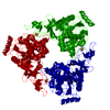
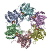
| Deposited unit | 
| ||||||||||||||||||
|---|---|---|---|---|---|---|---|---|---|---|---|---|---|---|---|---|---|---|---|
| 1 |
| ||||||||||||||||||
| 2 | 
| ||||||||||||||||||
| Unit cell |
| ||||||||||||||||||
| Components on special symmetry positions |
| ||||||||||||||||||
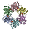
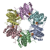
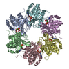
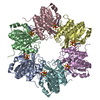
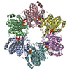
| Details | The biological assembly is a hexamer |
-Components
| #1: Protein | Mass: 23847.953 Da / Num. of mol.: 6 Source method: isolated from a genetically manipulated source Source: (gene. exp.) Pyrococcus horikoshii (archaea) / Strain: OT3 / Plasmid: pET11a / Production host:  #2: Chemical |   Mass: 35.453 Da / Num. of mol.: 3 / Source method: obtained synthetically / Formula: Cl Mass: 35.453 Da / Num. of mol.: 3 / Source method: obtained synthetically / Formula: Cl#3: Water | ChemComp-HOH / |  Mass: 18.015 Da / Num. of mol.: 663 / Source method: isolated from a natural source / Formula: H2O Mass: 18.015 Da / Num. of mol.: 663 / Source method: isolated from a natural source / Formula: H2OHas protein modification | Y | |
|---|
-Experimental details
-Experiment
| Experiment | Method: X-RAY DIFFRACTION / Number of used crystals: 1 |
|---|
- Sample preparation
| Crystal | Density Matthews: 2.11 Å3/Da / Density % sol: 41.63 % |
|---|---|
| Crystal grow | Temperature: 291 K / Method: microbatch / pH: 5.3 Details: 40%(v/v) 1,2-propanediol, Acetate pH 4.5, 0.05M Ca(OAc)2, pH 5.3, MICROBATCH, temperature 291K |
-Data collection
| Diffraction | Mean temperature: 100 K |
|---|---|
| Diffraction source | Source: SYNCHROTRON / Site: SPring-8  / Beamline: BL26B1 / Wavelength: 0.97907 Å / Beamline: BL26B1 / Wavelength: 0.97907 Å |
| Detector | Type: RIGAKU RAXIS V / Detector: IMAGE PLATE / Date: Apr 19, 2006 |
| Radiation | Protocol: SINGLE WAVELENGTH / Monochromatic (M) / Laue (L): M / Scattering type: x-ray |
| Radiation wavelength | Wavelength: 0.97907 Å / Relative weight: 1 |
| Reflection | Resolution: 2.3→30 Å / Num. all: 54164 / Num. obs: 54164 / % possible obs: 100 % / Observed criterion σ(F): 0 / Observed criterion σ(I): 0 / Redundancy: 7.4 % / Biso Wilson estimate: 30.846 Å2 / Rmerge(I) obs: 0.081 / Net I/σ(I): 20.8 |
| Reflection shell | Resolution: 2.3→2.38 Å / Redundancy: 7.4 % / Rmerge(I) obs: 0.148 / Mean I/σ(I) obs: 12.16 / % possible all: 100 |
- Processing
| Software |
| |||||||||||||||||||||||||
|---|---|---|---|---|---|---|---|---|---|---|---|---|---|---|---|---|---|---|---|---|---|---|---|---|---|---|
| Refinement | Method to determine structure: SAD / Resolution: 2.3→29.94 Å / Isotropic thermal model: RESTRAINED / Cross valid method: TROUGHOUT / σ(F): 0 / Stereochemistry target values: Engh & Huber
| |||||||||||||||||||||||||
| Displacement parameters | Biso mean: 31.8 Å2
| |||||||||||||||||||||||||
| Refine analyze |
| |||||||||||||||||||||||||
| Refinement step | Cycle: LAST / Resolution: 2.3→29.94 Å
| |||||||||||||||||||||||||
| Refine LS restraints |
| |||||||||||||||||||||||||
| LS refinement shell | Resolution: 2.3→2.4 Å / Rfactor Rfree error: 0.015
|
