 Movie
Movie Controller

[English] 日本語
 Yorodumi
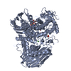
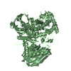
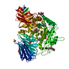
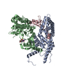
- PDB-2eae: Crystal structure of 1,2-a-L-fucosidase from Bifidobacterium bifi... -
+ Open data

- Basic information
| Entry | Database: PDB / ID: 2eae | |||||||||
|---|---|---|---|---|---|---|---|---|---|---|
| Title | Crystal structure of 1,2-a-L-fucosidase from Bifidobacterium bifidum in complexes with products | |||||||||
 Components Components | Alpha-fucosidase | |||||||||
 Keywords Keywords | HYDROLASE / fucosidase / glycoside hydrolase | |||||||||
| Function / homology |  Function and homology information Function and homology informationalpha-L-fucosidase activity / carbohydrate metabolic process / metal ion binding Similarity search - Function | |||||||||
| Biological species |  Bifidobacterium bifidum (bacteria) Bifidobacterium bifidum (bacteria) | |||||||||
| Method |  X-RAY DIFFRACTION / SYNCHROTRON / MOLECULAR REPLACEMENT / Resolution: 1.8 Å X-RAY DIFFRACTION / SYNCHROTRON / MOLECULAR REPLACEMENT / Resolution: 1.8 Å | |||||||||
 Authors Authors | Nagae, M. / Tsuchiya, A. / Katayama, T. / Yamamoto, K. / Wakatsuki, S. / Kato, R. | |||||||||
 Citation Citation | Journal: J.Biol.Chem. / Year: 2007 Title: Structural basis on the catalytic reaction mechanism of novel 1,2-alpha-L-fucosidase (AFCA) from Bifidobacterium bifidum Authors: Nagae, M. / Tsuchiya, A. / Katayama, T. / Yamamoto, K. / Wakatsuki, S. / Kato, R. | |||||||||
| History |
|
- Structure visualization
| Structure viewer | Molecule: MolmilJmol/JSmol |
|---|
- Downloads & links
-Download
| PDBx/mmCIF format | 2eae.cif.gz | 191.9 KB | Display | PDBx/mmCIF format |
|---|---|---|---|---|
| PDB format | pdb2eae.ent.gz | 149.8 KB | Display | PDB format |
| PDBx/mmJSON format | 2eae.json.gz | Tree view | PDBx/mmJSON format | |
| Others |  Other downloads Other downloads |
-Validation report
| Arichive directory | https://data.pdbj.org/pub/pdb/validation_reports/ea/2eaeftp://data.pdbj.org/pub/pdb/validation_reports/ea/2eae | HTTPS FTP |
|---|
-Related structure data
| Related structure data |  2eabC  2eacC  2eadC  2eaf C: citing same article ( |
|---|---|
| Similar structure data |


-Links
 PDBj
PDBj


- Assembly
| Deposited unit | 
| ||||||||
|---|---|---|---|---|---|---|---|---|---|
| 1 |
| ||||||||
| Unit cell |
|
-Components
-Protein , 1 types, 1 molecules A
| #1: Protein | Mass: 96795.992 Da / Num. of mol.: 1 / Fragment: catalytic domain / Mutation: D766A Source method: isolated from a genetically manipulated source Source: (gene. exp.) Bifidobacterium bifidum (bacteria) / Plasmid: pET3-a / Species (production host): Escherichia coli / Production host: |
|---|
-Sugars , 3 types, 3 molecules 
| #2: Polysaccharide | beta-D-galactopyranose-(1-4)-alpha-D-glucopyranose / alpha-lactose  Source method: isolated from a genetically manipulated source Details: oligosaccharide / References: alpha-lactose |
|---|---|
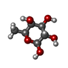
| #3: Sugar | ChemComp-FUC /  Type: L-saccharide, alpha linking / Mass: 164.156 Da / Num. of mol.: 1 Type: L-saccharide, alpha linking / Mass: 164.156 Da / Num. of mol.: 1Source method: isolated from a genetically manipulated source Formula: C6H12O5 |
| #4: Sugar | ChemComp-FUL /  Type: L-saccharide, beta linking / Mass: 164.156 Da / Num. of mol.: 1 Type: L-saccharide, beta linking / Mass: 164.156 Da / Num. of mol.: 1Source method: isolated from a genetically manipulated source Formula: C6H12O5 |
-Non-polymers , 2 types, 554 molecules 

| #5: Chemical |  Mass: 40.078 Da / Num. of mol.: 2 / Source method: obtained synthetically / Formula: Ca Mass: 40.078 Da / Num. of mol.: 2 / Source method: obtained synthetically / Formula: Ca#6: Water | ChemComp-HOH / | Mass: 18.015 Da / Num. of mol.: 552 / Source method: isolated from a natural source / Formula: H2O |
|---|
-Details
| Nonpolymer details | FUC AND FUL ARE IN ALTERNATE CONFORMATI |
|---|
-Experimental details
-Experiment
| Experiment | Method: X-RAY DIFFRACTION / Number of used crystals: 1 |
|---|
- Sample preparation
| Crystal | Density Matthews: 2.02 Å3/Da / Density % sol: 39.07 % |
|---|---|
| Crystal grow | Temperature: 293 K / Method: vapor diffusion, hanging drop / pH: 6 Details: 0.1M MES (pH6.0), 15% PEGMME2000, VAPOR DIFFUSION, HANGING DROP, temperature 293K |
-Data collection
| Diffraction | Mean temperature: 100 K |
|---|---|
| Diffraction source | Source: SYNCHROTRON / Site: Photon Factory  / Beamline: AR-NW12A / Wavelength: 1 Å / Beamline: AR-NW12A / Wavelength: 1 Å |
| Detector | Type: ADSC QUANTUM 210 / Detector: CCD / Date: Dec 17, 2005 |
| Radiation | Protocol: SINGLE WAVELENGTH / Monochromatic (M) / Laue (L): M / Scattering type: x-ray |
| Radiation wavelength | Wavelength: 1 Å / Relative weight: 1 |
| Reflection | Resolution: 1.8→171.5 Å / Num. all: 71461 / Num. obs: 69085 / % possible obs: 96.8 % / Redundancy: 12.1 % / Rsym value: 0.159 |
| Reflection shell | Resolution: 1.8→1.86 Å / Redundancy: 12.1 % / Num. unique all: 7005 / Rsym value: 0.383 / % possible all: 96.7 |
- Processing
| Software |
| ||||||||||||||||||||||||||||||||||||||||||||||||||||||||||||||||||||||||||||||||||||||||||||||||||||||||||||||||||||||||||||||||||||||||||||||||||||||||||||||||||||||||||
|---|---|---|---|---|---|---|---|---|---|---|---|---|---|---|---|---|---|---|---|---|---|---|---|---|---|---|---|---|---|---|---|---|---|---|---|---|---|---|---|---|---|---|---|---|---|---|---|---|---|---|---|---|---|---|---|---|---|---|---|---|---|---|---|---|---|---|---|---|---|---|---|---|---|---|---|---|---|---|---|---|---|---|---|---|---|---|---|---|---|---|---|---|---|---|---|---|---|---|---|---|---|---|---|---|---|---|---|---|---|---|---|---|---|---|---|---|---|---|---|---|---|---|---|---|---|---|---|---|---|---|---|---|---|---|---|---|---|---|---|---|---|---|---|---|---|---|---|---|---|---|---|---|---|---|---|---|---|---|---|---|---|---|---|---|---|---|---|---|---|---|---|
| Refinement | Method to determine structure: MOLECULAR REPLACEMENT / Resolution: 1.8→35.63 Å / Cor.coef. Fo:Fc: 0.959 / Cor.coef. Fo:Fc free: 0.942 / SU B: 2.945 / SU ML: 0.092 / Cross valid method: THROUGHOUT / ESU R: 0.145 / ESU R Free: 0.133 / Stereochemistry target values: MAXIMUM LIKELIHOOD
| ||||||||||||||||||||||||||||||||||||||||||||||||||||||||||||||||||||||||||||||||||||||||||||||||||||||||||||||||||||||||||||||||||||||||||||||||||||||||||||||||||||||||||
| Solvent computation | Ion probe radii: 0.8 Å / Shrinkage radii: 0.8 Å / VDW probe radii: 1.4 Å / Solvent model: MASK | ||||||||||||||||||||||||||||||||||||||||||||||||||||||||||||||||||||||||||||||||||||||||||||||||||||||||||||||||||||||||||||||||||||||||||||||||||||||||||||||||||||||||||
| Displacement parameters | Biso mean: 28.3 Å2
| ||||||||||||||||||||||||||||||||||||||||||||||||||||||||||||||||||||||||||||||||||||||||||||||||||||||||||||||||||||||||||||||||||||||||||||||||||||||||||||||||||||||||||
| Refinement step | Cycle: LAST / Resolution: 1.8→35.63 Å
| ||||||||||||||||||||||||||||||||||||||||||||||||||||||||||||||||||||||||||||||||||||||||||||||||||||||||||||||||||||||||||||||||||||||||||||||||||||||||||||||||||||||||||
| Refine LS restraints |
| ||||||||||||||||||||||||||||||||||||||||||||||||||||||||||||||||||||||||||||||||||||||||||||||||||||||||||||||||||||||||||||||||||||||||||||||||||||||||||||||||||||||||||
| LS refinement shell | Resolution: 1.797→1.844 Å / Total num. of bins used: 20
|
