 Movie
Movie Controller

[English] 日本語
 Yorodumi
- PDB-2e33: Structural basis for selection of glycosylated substrate by SCFFb... -
+ Open data

- Basic information
| Entry | Database: PDB / ID: 2.0E+33 | |||||||||
|---|---|---|---|---|---|---|---|---|---|---|
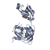
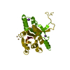
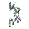
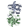
| Title | Structural basis for selection of glycosylated substrate by SCFFbs1 ubiquitin ligase | |||||||||
 Components Components |
| |||||||||
 Keywords Keywords | LIGASE/HYDROLASE / ubiquitin / SCF / Fbs1 / RNaseB / LIGASE-HYDROLASE COMPLEX | |||||||||
| Function / homology |  Function and homology information Function and homology informationextrinsic component of postsynaptic membrane / : / glycoprotein catabolic process / regulation of protein catabolic process at postsynapse, modulating synaptic transmission / Neddylation / Antigen processing: Ubiquitination & Proteasome degradation / pancreatic ribonuclease / ribonuclease A activity / SCF ubiquitin ligase complex / SCF-dependent proteasomal ubiquitin-dependent protein catabolic process ...extrinsic component of postsynaptic membrane / : / glycoprotein catabolic process / regulation of protein catabolic process at postsynapse, modulating synaptic transmission / Neddylation / Antigen processing: Ubiquitination & Proteasome degradation / pancreatic ribonuclease / ribonuclease A activity / SCF ubiquitin ligase complex / SCF-dependent proteasomal ubiquitin-dependent protein catabolic process / regulation of protein ubiquitination / RNA nuclease activity / ERAD pathway / amyloid-beta binding / carbohydrate binding / nucleic acid binding / dendritic spine / ubiquitin-dependent protein catabolic process / defense response to Gram-positive bacterium / protein ubiquitination / negative regulation of cell population proliferation / hydrolase activity / glutamatergic synapse / extracellular region / cytoplasm / cytosol Similarity search - Function | |||||||||
| Biological species |  | |||||||||
| Method |  X-RAY DIFFRACTION / SYNCHROTRON / MOLECULAR REPLACEMENT / Resolution: 2.7 Å X-RAY DIFFRACTION / SYNCHROTRON / MOLECULAR REPLACEMENT / Resolution: 2.7 Å | |||||||||
 Authors Authors | Mizushima, T. / Yoshida, Y. / Kumanomidou, T. / Hasegawa, Y. / Yamane, T. / Tanaka, K. | |||||||||
 Citation Citation | Journal: Proc.Natl.Acad.Sci.Usa / Year: 2007 Title: Structural basis for the selection of glycosylated substrates by SCFFbs1 ubiquitin ligase Authors: Mizushima, T. / Yoshida, Y. / Kumanomidou, T. / Hasegawa, Y. / Suzuki, A. / Yamane, T. / Tanaka, K. | |||||||||
| History |
|
- Structure visualization
| Structure viewer | Molecule: MolmilJmol/JSmol |
|---|
- Downloads & links
-Download
| PDBx/mmCIF format | 2e33.cif.gz | 76.1 KB | Display | PDBx/mmCIF format |
|---|---|---|---|---|
| PDB format | pdb2e33.ent.gz | 55.7 KB | Display | PDB format |
| PDBx/mmJSON format | 2e33.json.gz | Tree view | PDBx/mmJSON format | |
| Others |  Other downloads Other downloads |
-Validation report
| Arichive directory | https://data.pdbj.org/pub/pdb/validation_reports/e3/2e33ftp://data.pdbj.org/pub/pdb/validation_reports/e3/2e33 | HTTPS FTP |
|---|
-Related structure data
| Related structure data |  2e31C  2e32C  1umhS C: citing same article ( S: Starting model for refinement |
|---|---|
| Similar structure data |


-Links
 PDBj
PDBj


- Assembly
| Deposited unit | 
| ||||||||
|---|---|---|---|---|---|---|---|---|---|
| 1 |
| ||||||||
| Unit cell |
|
-Components
| #1: Protein | Mass: 22593.631 Da / Num. of mol.: 1 / Fragment: Residues 105-297 Source method: isolated from a genetically manipulated source Source: (gene. exp.)  |
|---|---|
| #2: Protein | Mass: 13708.326 Da / Num. of mol.: 1 / Source method: isolated from a natural source / Source: (natural) |
| #3: Polysaccharide | alpha-D-mannopyranose-(1-3)-[alpha-D-mannopyranose-(1-6)]beta-D-mannopyranose-(1-4)-2-acetamido-2- ...alpha-D-mannopyranose-(1-3)-[alpha-D-mannopyranose-(1-6)]beta-D-mannopyranose-(1-4)-2-acetamido-2-deoxy-beta-D-glucopyranose-(1-4)-2-acetamido-2-deoxy-beta-D-glucopyranose Source method: isolated from a genetically manipulated source |
| Has protein modification | Y |
-Experimental details
-Experiment
| Experiment | Method: X-RAY DIFFRACTION / Number of used crystals: 1 |
|---|
- Sample preparation
| Crystal | Density Matthews: 3.74 Å3/Da / Density % sol: 67.14 % |
|---|---|
| Crystal grow | Temperature: 293 K / Method: vapor diffusion, sitting drop / pH: 7.5 Details: 2.0% (v/v) PEG 400, 0.1M HEPES, 2.1M ammonium sulfate, pH 7.5, VAPOR DIFFUSION, SITTING DROP, temperature 293K |
-Data collection
| Diffraction | Mean temperature: 100 K |
|---|---|
| Diffraction source | Source: SYNCHROTRON / Site: SPring-8  / Beamline: BL44XU / Wavelength: 0.9 Å / Beamline: BL44XU / Wavelength: 0.9 Å |
| Detector | Type: Bruker DIP-6040 / Detector: CCD / Date: Jul 23, 2005 |
| Radiation | Protocol: SINGLE WAVELENGTH / Monochromatic (M) / Laue (L): M / Scattering type: x-ray |
| Radiation wavelength | Wavelength: 0.9 Å / Relative weight: 1 |
| Reflection | Resolution: 2.7→149.1 Å / Num. obs: 15839 / % possible obs: 99.4 % / Observed criterion σ(I): 0 / Redundancy: 11.8 % / Biso Wilson estimate: 62.6 Å2 / Rmerge(I) obs: 0.078 / Net I/σ(I): 13.9 |
| Reflection shell | Resolution: 2.7→2.8 Å / Redundancy: 6.4 % / Rmerge(I) obs: 0.325 / Mean I/σ(I) obs: 5.2 / % possible all: 97.9 |
- Processing
| Software |
| ||||||||||||||||||||||||||||||||||||||||||||||||||||||||||||||||||||||||||||||||||||||||||||||||||||||||||||||||||||||||||||||||||||||||||||||||||||||||||||||||||||||||||
|---|---|---|---|---|---|---|---|---|---|---|---|---|---|---|---|---|---|---|---|---|---|---|---|---|---|---|---|---|---|---|---|---|---|---|---|---|---|---|---|---|---|---|---|---|---|---|---|---|---|---|---|---|---|---|---|---|---|---|---|---|---|---|---|---|---|---|---|---|---|---|---|---|---|---|---|---|---|---|---|---|---|---|---|---|---|---|---|---|---|---|---|---|---|---|---|---|---|---|---|---|---|---|---|---|---|---|---|---|---|---|---|---|---|---|---|---|---|---|---|---|---|---|---|---|---|---|---|---|---|---|---|---|---|---|---|---|---|---|---|---|---|---|---|---|---|---|---|---|---|---|---|---|---|---|---|---|---|---|---|---|---|---|---|---|---|---|---|---|---|---|---|
| Refinement | Method to determine structure: MOLECULAR REPLACEMENT Starting model: PDB ENTRY 1UMH Resolution: 2.7→50 Å / Cor.coef. Fo:Fc: 0.931 / Cor.coef. Fo:Fc free: 0.881 / SU B: 11.824 / SU ML: 0.244 / Cross valid method: THROUGHOUT / ESU R: 0.4 / ESU R Free: 0.318 / Stereochemistry target values: MAXIMUM LIKELIHOOD / Details: HYDROGENS HAVE BEEN ADDED IN THE RIDING POSITIONS
| ||||||||||||||||||||||||||||||||||||||||||||||||||||||||||||||||||||||||||||||||||||||||||||||||||||||||||||||||||||||||||||||||||||||||||||||||||||||||||||||||||||||||||
| Solvent computation | Ion probe radii: 0.8 Å / Shrinkage radii: 0.8 Å / VDW probe radii: 1.2 Å / Solvent model: MASK | ||||||||||||||||||||||||||||||||||||||||||||||||||||||||||||||||||||||||||||||||||||||||||||||||||||||||||||||||||||||||||||||||||||||||||||||||||||||||||||||||||||||||||
| Displacement parameters | Biso mean: 54.8 Å2 | ||||||||||||||||||||||||||||||||||||||||||||||||||||||||||||||||||||||||||||||||||||||||||||||||||||||||||||||||||||||||||||||||||||||||||||||||||||||||||||||||||||||||||
| Refinement step | Cycle: LAST / Resolution: 2.7→50 Å
| ||||||||||||||||||||||||||||||||||||||||||||||||||||||||||||||||||||||||||||||||||||||||||||||||||||||||||||||||||||||||||||||||||||||||||||||||||||||||||||||||||||||||||
| Refine LS restraints |
| ||||||||||||||||||||||||||||||||||||||||||||||||||||||||||||||||||||||||||||||||||||||||||||||||||||||||||||||||||||||||||||||||||||||||||||||||||||||||||||||||||||||||||
| LS refinement shell | Resolution: 2.7→2.77 Å / Total num. of bins used: 20
|
