 Movie
Movie Controller

[English] 日本語
 Yorodumi
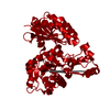
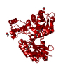
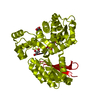
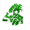
- PDB-2e20: Crystal structure of Salmonella typhimurium propionate kinase (Td... -
+ Open data

- Basic information
| Entry | Database: PDB / ID: 2.0E+20 | ||||||
|---|---|---|---|---|---|---|---|
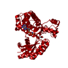
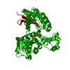
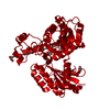
| Title | Crystal structure of Salmonella typhimurium propionate kinase (TdcD) in complex with diadenosine tetraphosphate (Ap4A) | ||||||
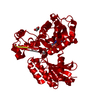
 Components Components | Propionate Kinase | ||||||
 Keywords Keywords | TRANSFERASE / Propionate kinase / TdcD / Native / Acetate kinase / Nucleotide / Ap4A / ADP / ATP / AMPPNP | ||||||
| Function / homology |  Function and homology information Function and homology informationpropionate kinase / propionate kinase activity / : / acetate kinase activity / acetate metabolic process / ATP binding / metal ion binding / cytosol Similarity search - Function | ||||||
| Biological species |  Salmonella typhimurium (bacteria) Salmonella typhimurium (bacteria) | ||||||
| Method |  X-RAY DIFFRACTION / FOURIER SYNTHESIS / Resolution: 2.4 Å X-RAY DIFFRACTION / FOURIER SYNTHESIS / Resolution: 2.4 Å | ||||||
 Authors Authors | Simanshu, D.K. / Savithri, H.S. / Murthy, M.R.N. | ||||||
 Citation Citation | Journal: Proteins / Year: 2008 Title: Crystal structures of Salmonella typhimurium propionate kinase and its complex with Ap4A: evidence for a novel Ap4A synthetic activity. Authors: Simanshu, D.K. / Savithri, H.S. / Murthy, M.R.N. #1: Journal: J.Mol.Biol. / Year: 2005Title: Crystal structures of ADP and AMPPNP-bound propionate kinase (TdcD) from Salmonella typhimurium: comparison with members of acetate and sugar kinase/heat shock cognate 70/actin superfamily Authors: Simanshu, D.K. / Savithri, H.S. / Murthy, M.R. #2: Journal: ACTA CRYSTALLOGR.,SECT.F / Year: 2005 Title: Cloning, expression, purification, crystallization and preliminary X-ray diffraction analysis of propionate kinase (TdcD) from Salmonella typhimurium Authors: Simanshu, D.K. / Murthy, M.R. | ||||||
| History |
|
- Structure visualization
| Structure viewer | Molecule: MolmilJmol/JSmol |
|---|
- Downloads & links
-Download
| PDBx/mmCIF format | 2e20.cif.gz | 93.5 KB | Display | PDBx/mmCIF format |
|---|---|---|---|---|
| PDB format | pdb2e20.ent.gz | 68.5 KB | Display | PDB format |
| PDBx/mmJSON format | 2e20.json.gz | Tree view | PDBx/mmJSON format | |
| Others |  Other downloads Other downloads |
-Validation report
| Arichive directory | https://data.pdbj.org/pub/pdb/validation_reports/e2/2e20ftp://data.pdbj.org/pub/pdb/validation_reports/e2/2e20 | HTTPS FTP |
|---|
-Related structure data
| Related structure data |  2e1yC 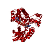 2e1zC  1x3mS S: Starting model for refinement C: citing same article ( |
|---|---|
| Similar structure data |


-Links
 PDBj
PDBj- Assembly
| Deposited unit | 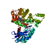
| |||||||||
|---|---|---|---|---|---|---|---|---|---|---|
| 1 | 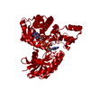
| |||||||||
| Unit cell |
| |||||||||
| Components on special symmetry positions |
|
-Components
| #1: Protein | Mass: 45246.398 Da / Num. of mol.: 1 Source method: isolated from a genetically manipulated source Source: (gene. exp.) Salmonella typhimurium (bacteria) / Strain: IFO 12529 / Gene: TdcD / Plasmid: pRSET C / Production host: |
|---|---|
| #2: Chemical | ChemComp-B4P / 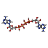  Mass: 836.387 Da / Num. of mol.: 1 / Source method: obtained synthetically / Formula: C20H28N10O19P4 Mass: 836.387 Da / Num. of mol.: 1 / Source method: obtained synthetically / Formula: C20H28N10O19P4 |
| #3: Chemical | ChemComp-EDO /   Mass: 62.068 Da / Num. of mol.: 1 / Source method: obtained synthetically / Formula: C2H6O2 Mass: 62.068 Da / Num. of mol.: 1 / Source method: obtained synthetically / Formula: C2H6O2 |
| #4: Water | ChemComp-HOH /  Mass: 18.015 Da / Num. of mol.: 152 / Source method: isolated from a natural source / Formula: H2O Mass: 18.015 Da / Num. of mol.: 152 / Source method: isolated from a natural source / Formula: H2O |
-Experimental details
-Experiment
| Experiment | Method: X-RAY DIFFRACTION / Number of used crystals: 1 |
|---|
- Sample preparation
| Crystal | Density Matthews: 2.6 Å3/Da / Density % sol: 52.63 % |
|---|---|
| Crystal grow | Temperature: 293 K / Method: vapor diffusion, hanging drop / pH: 6.5 Details: 0.1M Bis-Tris pH 6.5, 45% (v/v) pentaerythritol ethoxylate (15/4 EO/OH), 100mM ammonium sulfate, VAPOR DIFFUSION, HANGING DROP, temperature 293K |
-Data collection
| Diffraction | Mean temperature: 100 K |
|---|---|
| Diffraction source | Source: ROTATING ANODE / Type: RIGAKU RU200 / Wavelength: 1.5418 Å |
| Detector | Type: MAR scanner 345 mm plate / Detector: IMAGE PLATE / Date: Mar 23, 2006 / Details: Osmic mirror |
| Radiation | Monochromator: Mirror / Protocol: SINGLE WAVELENGTH / Monochromatic (M) / Laue (L): M / Scattering type: x-ray |
| Radiation wavelength | Wavelength: 1.5418 Å / Relative weight: 1 |
| Reflection | Resolution: 2.4→30 Å / Num. obs: 18755 / % possible obs: 99.2 % / Redundancy: 31.48 % / Biso Wilson estimate: 58.02 Å2 / Rmerge(I) obs: 0.074 / Net I/σ(I): 22.1 |
| Reflection shell | Resolution: 2.4→2.49 Å / Rmerge(I) obs: 0.413 / Mean I/σ(I) obs: 3.05 / Num. unique all: 1775 / % possible all: 95.2 |
- Processing
| Software |
| ||||||||||||||||||||||||||||||||||||||||||||||||||||||||||||||||||||||||||||||||||||||||||||||||||||||||||||||||||||||||||||||||||||||||||||||||||||||||||||||||||||||||||
|---|---|---|---|---|---|---|---|---|---|---|---|---|---|---|---|---|---|---|---|---|---|---|---|---|---|---|---|---|---|---|---|---|---|---|---|---|---|---|---|---|---|---|---|---|---|---|---|---|---|---|---|---|---|---|---|---|---|---|---|---|---|---|---|---|---|---|---|---|---|---|---|---|---|---|---|---|---|---|---|---|---|---|---|---|---|---|---|---|---|---|---|---|---|---|---|---|---|---|---|---|---|---|---|---|---|---|---|---|---|---|---|---|---|---|---|---|---|---|---|---|---|---|---|---|---|---|---|---|---|---|---|---|---|---|---|---|---|---|---|---|---|---|---|---|---|---|---|---|---|---|---|---|---|---|---|---|---|---|---|---|---|---|---|---|---|---|---|---|---|---|---|
| Refinement | Method to determine structure: FOURIER SYNTHESIS Starting model: PDB entry 1X3M Resolution: 2.4→30 Å / Cor.coef. Fo:Fc: 0.949 / Cor.coef. Fo:Fc free: 0.933 / SU B: 8.096 / SU ML: 0.19 / Cross valid method: THROUGHOUT / ESU R: 0.388 / ESU R Free: 0.261 / Stereochemistry target values: MAXIMUM LIKELIHOOD / Details: HYDROGENS HAVE BEEN ADDED IN THE RIDING POSITIONS
| ||||||||||||||||||||||||||||||||||||||||||||||||||||||||||||||||||||||||||||||||||||||||||||||||||||||||||||||||||||||||||||||||||||||||||||||||||||||||||||||||||||||||||
| Solvent computation | Ion probe radii: 0.8 Å / Shrinkage radii: 0.8 Å / VDW probe radii: 1.4 Å / Solvent model: MASK | ||||||||||||||||||||||||||||||||||||||||||||||||||||||||||||||||||||||||||||||||||||||||||||||||||||||||||||||||||||||||||||||||||||||||||||||||||||||||||||||||||||||||||
| Displacement parameters | Biso mean: 38.022 Å2
| ||||||||||||||||||||||||||||||||||||||||||||||||||||||||||||||||||||||||||||||||||||||||||||||||||||||||||||||||||||||||||||||||||||||||||||||||||||||||||||||||||||||||||
| Refinement step | Cycle: LAST / Resolution: 2.4→30 Å
| ||||||||||||||||||||||||||||||||||||||||||||||||||||||||||||||||||||||||||||||||||||||||||||||||||||||||||||||||||||||||||||||||||||||||||||||||||||||||||||||||||||||||||
| Refine LS restraints |
| ||||||||||||||||||||||||||||||||||||||||||||||||||||||||||||||||||||||||||||||||||||||||||||||||||||||||||||||||||||||||||||||||||||||||||||||||||||||||||||||||||||||||||
| LS refinement shell | Resolution: 2.4→2.462 Å / Total num. of bins used: 20
|
