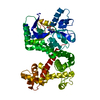
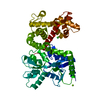
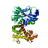
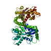
- PDB-2dx1: Crystal structure of RhoGEF protein Asef -
+
Open data
ID or keywords:
Loading...
-
Basic information
Entry
Database: PDB / ID: 2dx1
Title
Crystal structure of RhoGEF protein Asef
Components
Rho guanine nucleotide exchange factor 4
Keywords
SIGNALING PROTEIN / Rho-GEF / Structural Genomics / NPPSFA / National Project on Protein Structural and Functional Analyses / RIKEN Structural Genomics/Proteomics Initiative / RSGI
Function / homology
Function and homology information
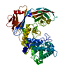
filopodium assembly / regulation of small GTPase mediated signal transduction / lamellipodium assembly / NRAGE signals death through JNK / CDC42 GTPase cycle / RHOA GTPase cycle / RAC1 GTPase cycle / guanyl-nucleotide exchange factor activity / ruffle membrane / G alpha (12/13) signalling events ...filopodium assembly / regulation of small GTPase mediated signal transduction / lamellipodium assembly / NRAGE signals death through JNK / CDC42 GTPase cycle / RHOA GTPase cycle / RAC1 GTPase cycle / guanyl-nucleotide exchange factor activity / ruffle membrane / G alpha (12/13) signalling events / intracellular signal transduction / protein domain specific binding / cytosol Similarity search - Function
In the structure databanks used in Yorodumi, some data are registered as the other names, "COVID-19 virus" and "2019-nCoV". Here are the details of the virus and the list of structure data.
Jan 31, 2019. EMDB accession codes are about to change! (news from PDBe EMDB page)
EMDB accession codes are about to change! (news from PDBe EMDB page)
The allocation of 4 digits for EMDB accession codes will soon come to an end. Whilst these codes will remain in use, new EMDB accession codes will include an additional digit and will expand incrementally as the available range of codes is exhausted. The current 4-digit format prefixed with “EMD-” (i.e. EMD-XXXX) will advance to a 5-digit format (i.e. EMD-XXXXX), and so on. It is currently estimated that the 4-digit codes will be depleted around Spring 2019, at which point the 5-digit format will come into force.
The EM Navigator/Yorodumi systems omit the EMD- prefix.
Related info.:Q: What is EMD? / ID/Accession-code notation in Yorodumi/EM Navigator
Yorodumi is a browser for structure data from EMDB, PDB, SASBDB, etc.
This page is also the successor to EM Navigator detail page, and also detail information page/front-end page for Omokage search.
The word "yorodu" (or yorozu) is an old Japanese word meaning "ten thousand". "mi" (miru) is to see.
Related info.:EMDB / PDB / SASBDB / Comparison of 3 databanks / Yorodumi Search / Aug 31, 2016. New EM Navigator & Yorodumi / Yorodumi Papers / Jmol/JSmol / Function and homology information / Changes in new EM Navigator and Yorodumi
 Movie
Movie Controller


 Open data
 Basic information
 Components
 Keywords
 Function and homology information
 Homo sapiens (human)
 X-RAY DIFFRACTION /
  Authors
 Citation
 Structure visualization
 Downloads & links
 Other downloads
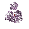


 PDBj
PDBj


 Assembly
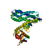
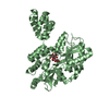
 Mass: 18.015 Da / Num. of mol.: 64 / Source method: isolated from a natural source / Formula: H2O
 Sample preparation
 / Beamline: BL44B2 / Wavelength: 0.9789, 0.9794, 0.9640
 Processing