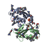
- PDB-2dx0: Crystal structure of the N-terminal SH2 domain of mouse phospholi... -
+
Open data
ID or keywords:
Loading...
-
Basic information
Entry
Database: PDB / ID: 2dx0
Title
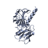
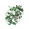
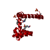
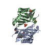
Crystal structure of the N-terminal SH2 domain of mouse phospholipase C-gamma 2
Components
Phospholipase C, gamma 2
Keywords
HYDROLASE / PHOSPHORIC DIESTER HYDROLASE / Structural Genomics / NPPSFA / National Project on Protein Structural and Functional Analyses / RIKEN Structural Genomics/Proteomics Initiative / RSGI
Function / homology
Function and homology information
Dectin-2 family / Toll Like Receptor 4 (TLR4) Cascade / inositol trisphosphate biosynthetic process / phosphatidylcholine phospholipase C activity / Synthesis of IP3 and IP4 in the cytosol / GPVI-mediated activation cascade / FCERI mediated MAPK activation / regulation of calcineurin-NFAT signaling cascade / Generation of second messenger molecules / Role of phospholipids in phagocytosis ...Dectin-2 family / Toll Like Receptor 4 (TLR4) Cascade / inositol trisphosphate biosynthetic process / phosphatidylcholine phospholipase C activity / Synthesis of IP3 and IP4 in the cytosol / GPVI-mediated activation cascade / FCERI mediated MAPK activation / regulation of calcineurin-NFAT signaling cascade / Generation of second messenger molecules / Role of phospholipids in phagocytosis / follicular B cell differentiation / positive regulation of dendritic cell cytokine production / phosphoinositide phospholipase C / FCERI mediated Ca+2 mobilization / antifungal innate immune response / cellular response to lectin / positive regulation of interleukin-23 production / Antigen activates B Cell Receptor (BCR) leading to generation of second messengers / CLEC7A (Dectin-1) signaling / phosphorylation-dependent protein binding / phosphatidylinositol metabolic process / positive regulation of cell cycle G1/S phase transition / response to yeast / phosphatidylinositol-4,5-bisphosphate phospholipase C activity / cell activation / C-type glycerophospholipase activity / positive regulation of phagocytosis, engulfment / phosphatidylinositol biosynthetic process / macrophage activation involved in immune response / phospholipid catabolic process / cellular response to lipid / DAP12 signaling / regulation of canonical NF-kappaB signal transduction / positive regulation of macrophage cytokine production / negative regulation of programmed cell death / positive regulation of neuroinflammatory response / toll-like receptor signaling pathway / stimulatory C-type lectin receptor signaling pathway / response to ATP / intracellular vesicle / phosphatidylinositol-mediated signaling / positive regulation of reactive oxygen species biosynthetic process / positive regulation of NLRP3 inflammasome complex assembly / regulation of lipid metabolic process / positive regulation of intracellular signal transduction / B cell activation / positive regulation of epithelial cell migration / positive regulation of interleukin-10 production / positive regulation of receptor internalization / response to magnesium ion / response to axon injury / positive regulation of interleukin-2 production / positive regulation of type I interferon production / phosphotyrosine residue binding / release of sequestered calcium ion into cytosol / positive regulation of interleukin-12 production / positive regulation of calcium-mediated signaling / lipopolysaccharide-mediated signaling pathway / cellular response to calcium ion / B cell differentiation / protein tyrosine kinase binding / B cell receptor signaling pathway / calcium-mediated signaling / positive regulation of interleukin-6 production / ruffle membrane / positive regulation of tumor necrosis factor production / T cell receptor signaling pathway / positive regulation of cytosolic calcium ion concentration / regulation of gene expression / scaffold protein binding / response to lipopolysaccharide / positive regulation of canonical NF-kappaB signal transduction / positive regulation of MAPK cascade / intracellular signal transduction / membrane raft / positive regulation of gene expression / protein kinase binding / perinuclear region of cytoplasm / plasma membrane / cytoplasm / cytosol Similarity search - Function
In the structure databanks used in Yorodumi, some data are registered as the other names, "COVID-19 virus" and "2019-nCoV". Here are the details of the virus and the list of structure data.
Jan 31, 2019. EMDB accession codes are about to change! (news from PDBe EMDB page)
EMDB accession codes are about to change! (news from PDBe EMDB page)
The allocation of 4 digits for EMDB accession codes will soon come to an end. Whilst these codes will remain in use, new EMDB accession codes will include an additional digit and will expand incrementally as the available range of codes is exhausted. The current 4-digit format prefixed with “EMD-” (i.e. EMD-XXXX) will advance to a 5-digit format (i.e. EMD-XXXXX), and so on. It is currently estimated that the 4-digit codes will be depleted around Spring 2019, at which point the 5-digit format will come into force.
The EM Navigator/Yorodumi systems omit the EMD- prefix.
Related info.:Q: What is EMD? / ID/Accession-code notation in Yorodumi/EM Navigator
Yorodumi is a browser for structure data from EMDB, PDB, SASBDB, etc.
This page is also the successor to EM Navigator detail page, and also detail information page/front-end page for Omokage search.
The word "yorodu" (or yorozu) is an old Japanese word meaning "ten thousand". "mi" (miru) is to see.
Related info.:EMDB / PDB / SASBDB / Comparison of 3 databanks / Yorodumi Search / Aug 31, 2016. New EM Navigator & Yorodumi / Yorodumi Papers / Jmol/JSmol / Function and homology information / Changes in new EM Navigator and Yorodumi
 Movie
Movie Controller

 Yorodumi
 Open data
 Basic information
 Components
 Keywords
 Function and homology information
 X-RAY DIFFRACTION /
  Authors
 Citation
 Structure visualization
 Downloads & links
 Other downloads


 PDBj
PDBj


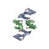
 Assembly
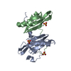


 Mass: 96.063 Da / Num. of mol.: 3 / Source method: obtained synthetically / Formula: SO4
 Mass: 18.015 Da / Num. of mol.: 102 / Source method: isolated from a natural source / Formula: H2O
 Sample preparation
 / Beamline: BL26B2 / Wavelength: 0.9791, 0.9794, 0.9700
 Processing