 Movie
Movie Controller


+ Open data

- Basic information
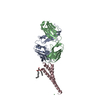
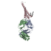
| Entry | Database: PDB / ID: 2dwe | ||||||
|---|---|---|---|---|---|---|---|
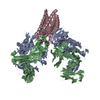
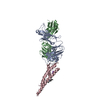
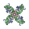
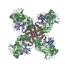
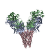
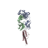
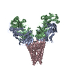
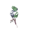
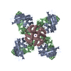
| Title | Crystal structure of KcsA-FAB-TBA complex in Rb+ | ||||||
 Components Components |
| ||||||
 Keywords Keywords | MEMBRANE PROTEIN / POTASSIUM CHANNEL / TETRABUTYLAMMONIUM / K+ / KcsA | ||||||
| Function / homology |  Function and homology information Function and homology informationaction potential / voltage-gated potassium channel activity / voltage-gated potassium channel complex / identical protein binding Similarity search - Function | ||||||
| Biological species |  Streptomyces lividans (bacteria) Streptomyces lividans (bacteria) | ||||||
| Method |  X-RAY DIFFRACTION / SYNCHROTRON / MOLECULAR REPLACEMENT / Resolution: 2.5 Å X-RAY DIFFRACTION / SYNCHROTRON / MOLECULAR REPLACEMENT / Resolution: 2.5 Å | ||||||
 Authors Authors | Yohannan, S. / Zhou, Y. | ||||||
 Citation Citation | Journal: J.Mol.Biol. / Year: 2007 Title: Crystallographic Study of the Tetrabutylammonium Block to the KcsA K(+) Channel Authors: Yohannan, S. / Hu, Y. / Zhou, Y. | ||||||
| History |
|
- Structure visualization
| Structure viewer | Molecule: MolmilJmol/JSmol |
|---|
- Downloads & links
-Download
| PDBx/mmCIF format | 2dwe.cif.gz | 121.3 KB | Display | PDBx/mmCIF format |
|---|---|---|---|---|
| PDB format | pdb2dwe.ent.gz | 92 KB | Display | PDB format |
| PDBx/mmJSON format | 2dwe.json.gz | Tree view | PDBx/mmJSON format | |
| Others |  Other downloads Other downloads |
-Validation report
| Summary document | 2dwe_validation.pdf.gz | 715.2 KB | Display | wwPDB validaton report |
|---|---|---|---|---|
| Full document | 2dwe_full_validation.pdf.gz | 735.6 KB | Display | |
| Data in XML | 2dwe_validation.xml.gz | 25 KB | Display | |
| Data in CIF | 2dwe_validation.cif.gz | 34 KB | Display | |
| Arichive directory | https://data.pdbj.org/pub/pdb/validation_reports/dw/2dweftp://data.pdbj.org/pub/pdb/validation_reports/dw/2dwe | HTTPS FTP |
-Related structure data
| Related structure data |  2dwdC  2hvjC 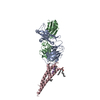 2hvkC  1k4cS S: Starting model for refinement C: citing same article ( |
|---|---|
| Similar structure data |


-Links
 PDBj
PDBj


- Assembly
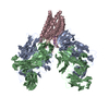
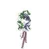
| Deposited unit | 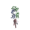
| |||||||||||||||
|---|---|---|---|---|---|---|---|---|---|---|---|---|---|---|---|---|
| 1 | 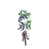
| |||||||||||||||
| Unit cell |
| |||||||||||||||
| Components on special symmetry positions |
| |||||||||||||||
| Details | biological assembly is a tetramer generated by: chain A, chain B (x,y,z (1_555),2-x,2-y,z (2_775),2-y,x,z (3_755),y,2-x,z (4_575)); chain C (x,y,z (1_555),2-x,2-y,z (2_775),2-y,x,z (3_755),y,2-x,z (4_575)) |
-Components
-Protein , 1 types, 1 molecules C
| #3: Protein | Mass: 10978.736 Da / Num. of mol.: 1 / Fragment: residues 22-124 / Mutation: L90C Source method: isolated from a genetically manipulated source Source: (gene. exp.) Streptomyces lividans (bacteria) / Plasmid: pQE60 / Production host: |
|---|
-Antibody , 2 types, 2 molecules AB
| #1: Antibody | Mass: 23411.242 Da / Num. of mol.: 1 / Source method: isolated from a natural source / Details: hybridoma cell line / Source: (natural) |
|---|---|
| #2: Antibody | Mass: 23435.738 Da / Num. of mol.: 1 / Source method: isolated from a natural source / Details: hybridoma cell line / Source: (natural) |
-Non-polymers , 5 types, 103 molecules 


| #4: Chemical |  Mass: 85.468 Da / Num. of mol.: 3 / Source method: obtained synthetically / Formula: Rb Mass: 85.468 Da / Num. of mol.: 3 / Source method: obtained synthetically / Formula: Rb#5: Chemical | ChemComp-L2C / ( |  Mass: 414.619 Da / Num. of mol.: 1 / Source method: obtained synthetically / Formula: C24H46O5 Mass: 414.619 Da / Num. of mol.: 1 / Source method: obtained synthetically / Formula: C24H46O5#6: Chemical | ChemComp-F09 / |  Mass: 144.254 Da / Num. of mol.: 1 / Source method: obtained synthetically / Formula: C9H20O Mass: 144.254 Da / Num. of mol.: 1 / Source method: obtained synthetically / Formula: C9H20O#7: Chemical | ChemComp-TBA / |  Mass: 242.464 Da / Num. of mol.: 1 / Source method: obtained synthetically / Formula: C16H36N Mass: 242.464 Da / Num. of mol.: 1 / Source method: obtained synthetically / Formula: C16H36N#8: Water | ChemComp-HOH / | Mass: 18.015 Da / Num. of mol.: 97 / Source method: isolated from a natural source / Formula: H2O |
|---|
-Details
| Has protein modification | Y |
|---|
-Experimental details
-Experiment
| Experiment | Method: X-RAY DIFFRACTION / Number of used crystals: 1 |
|---|
- Sample preparation
| Crystal | Density Matthews: 3.91 Å3/Da / Density % sol: 68.57 % |
|---|---|
| Crystal grow | Temperature: 293 K / Method: vapor diffusion, sitting drop / pH: 6.5 Details: 18-25% PEG 400, 50mM MG(AC)2, 50mM NaAc(pH 5) or Na cacodylate(pH 6) or HEPES(pH 7), pH 6.5, VAPOR DIFFUSION, SITTING DROP, temperature 293K |
-Data collection
| Diffraction | Mean temperature: 100 K |
|---|---|
| Diffraction source | Source: SYNCHROTRON / Site: NSLS  / Beamline: X25 / Wavelength: 1.1 Å / Beamline: X25 / Wavelength: 1.1 Å |
| Detector | Type: ADSC QUANTUM 315 / Detector: CCD / Date: Oct 1, 2005 |
| Radiation | Monochromator: Si-111 double crystal / Protocol: SINGLE WAVELENGTH / Monochromatic (M) / Laue (L): M / Scattering type: x-ray |
| Radiation wavelength | Wavelength: 1.1 Å / Relative weight: 1 |
| Reflection | Resolution: 2.5→26.84 Å / Num. all: 30997 / Num. obs: 30341 / % possible obs: 97.9 % / Observed criterion σ(F): 0 / Observed criterion σ(I): 0 / Redundancy: 4.2 % / Rmerge(I) obs: 0.06 |
| Reflection shell | Resolution: 2.5→2.59 Å / Redundancy: 4.2 % / % possible all: 99.1 |
- Processing
| Software |
| ||||||||||||||||||||
|---|---|---|---|---|---|---|---|---|---|---|---|---|---|---|---|---|---|---|---|---|---|
| Refinement | Method to determine structure: MOLECULAR REPLACEMENT Starting model: PDB entry 1K4C Resolution: 2.5→26.84 Å / Cross valid method: THROUGHOUT / σ(F): 0 / Stereochemistry target values: Engh & Huber
| ||||||||||||||||||||
| Displacement parameters |
| ||||||||||||||||||||
| Refinement step | Cycle: LAST / Resolution: 2.5→26.84 Å
| ||||||||||||||||||||
| Refine LS restraints |
|
