 Movie
Movie Controller

[English] 日本語
 Yorodumi
- PDB-2dw6: Crystal structure of the mutant K184A of D-Tartrate Dehydratase f... -
+ Open data

- Basic information
| Entry | Database: PDB / ID: 2dw6 | ||||||
|---|---|---|---|---|---|---|---|
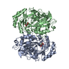
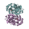
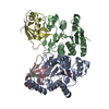
| Title | Crystal structure of the mutant K184A of D-Tartrate Dehydratase from Bradyrhizobium japonicum complexed with Mg++ and D-tartrate | ||||||
 Components Components | Bll6730 protein | ||||||
 Keywords Keywords | LYASE / D-Tartrate Dehydratase / Enolase superfamily / D-tartrate / L-tartrate | ||||||
| Function / homology |  Function and homology information Function and homology informationD(-)-tartrate dehydratase / D(-)-tartrate dehydratase activity / D-galactonate catabolic process / hydro-lyase activity / protein homooligomerization / magnesium ion binding Similarity search - Function | ||||||
| Biological species |  Bradyrhizobium japonicum (bacteria) Bradyrhizobium japonicum (bacteria) | ||||||
| Method |  X-RAY DIFFRACTION / SYNCHROTRON / MOLECULAR REPLACEMENT / Resolution: 2.3 Å X-RAY DIFFRACTION / SYNCHROTRON / MOLECULAR REPLACEMENT / Resolution: 2.3 Å | ||||||
 Authors Authors | Fedorov, A.A. / Fedorov, E.V. / Yew, W.S. / Wood, B.M. / Gerlt, J.A. / Almo, S.C. | ||||||
 Citation Citation | Journal: Biochemistry / Year: 2006 Title: Evolution of Enzymatic Activities in the Enolase Superfamily: d-Tartrate Dehydratase from Bradyrhizobium japonicum Authors: Yew, W.S. / Fedorov, A.A. / Fedorov, E.V. / Wood, B.M. / Almo, S.C. / Gerlt, J.A. | ||||||
| History |
|
- Structure visualization
| Structure viewer | Molecule: MolmilJmol/JSmol |
|---|
- Downloads & links
-Download
| PDBx/mmCIF format | 2dw6.cif.gz | 311.7 KB | Display | PDBx/mmCIF format |
|---|---|---|---|---|
| PDB format | pdb2dw6.ent.gz | 253.5 KB | Display | PDB format |
| PDBx/mmJSON format | 2dw6.json.gz | Tree view | PDBx/mmJSON format | |
| Others |  Other downloads Other downloads |
-Validation report
| Summary document | 2dw6_validation.pdf.gz | 491.8 KB | Display | wwPDB validaton report |
|---|---|---|---|---|
| Full document | 2dw6_full_validation.pdf.gz | 516.9 KB | Display | |
| Data in XML | 2dw6_validation.xml.gz | 59.4 KB | Display | |
| Data in CIF | 2dw6_validation.cif.gz | 82.8 KB | Display | |
| Arichive directory | https://data.pdbj.org/pub/pdb/validation_reports/dw/2dw6ftp://data.pdbj.org/pub/pdb/validation_reports/dw/2dw6 | HTTPS FTP |
-Related structure data
| Related structure data |  2dw7C 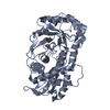 1tzzS S: Starting model for refinement C: citing same article ( |
|---|---|
| Similar structure data |


-Links
 PDBj
PDBj


- Assembly
| Deposited unit | 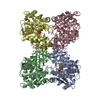
| ||||||||
|---|---|---|---|---|---|---|---|---|---|
| 1 | 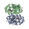
| ||||||||
| 2 | 
| ||||||||
| Unit cell |
| ||||||||
| Details | The biological assembly is a dimer. There are two dimers in the asymmetric unit. |
-Components
| #1: Protein | Mass: 43053.008 Da / Num. of mol.: 4 / Mutation: K184A Source method: isolated from a genetically manipulated source Source: (gene. exp.) Bradyrhizobium japonicum (bacteria) / Plasmid: pET-15b / Species (production host): Escherichia coli / Production host: #2: Chemical | ChemComp-MG /   Mass: 24.305 Da / Num. of mol.: 4 / Source method: obtained synthetically / Formula: Mg Mass: 24.305 Da / Num. of mol.: 4 / Source method: obtained synthetically / Formula: Mg#3: Chemical |   Mass: 150.087 Da / Num. of mol.: 3 / Source method: obtained synthetically / Formula: C4H6O6 Mass: 150.087 Da / Num. of mol.: 3 / Source method: obtained synthetically / Formula: C4H6O6#4: Chemical | ChemComp-TLA / |   Mass: 150.087 Da / Num. of mol.: 1 / Source method: obtained synthetically / Formula: C4H6O6 Mass: 150.087 Da / Num. of mol.: 1 / Source method: obtained synthetically / Formula: C4H6O6#5: Water | ChemComp-HOH / |  Mass: 18.015 Da / Num. of mol.: 466 / Source method: isolated from a natural source / Formula: H2O Mass: 18.015 Da / Num. of mol.: 466 / Source method: isolated from a natural source / Formula: H2O |
|---|
-Experimental details
-Experiment
| Experiment | Method: X-RAY DIFFRACTION / Number of used crystals: 1 |
|---|
- Sample preparation
| Crystal | Density Matthews: 4.24 Å3/Da / Density % sol: 71 % |
|---|---|
| Crystal grow | Temperature: 293 K / Method: vapor diffusion, hanging drop / pH: 8.5 Details: 0.8M K/Na L-tartrate, 0.1M Tris.HCl, 0.5% PEG MME 5000, pH 8.5, VAPOR DIFFUSION, HANGING DROP, temperature 293.0K |
-Data collection
| Diffraction | Mean temperature: 100 K |
|---|---|
| Diffraction source | Source: SYNCHROTRON / Site: NSLS  / Beamline: X4A / Wavelength: 0.97911 Å / Beamline: X4A / Wavelength: 0.97911 Å |
| Detector | Type: ADSC QUANTUM 4 / Detector: CCD / Date: Apr 13, 2006 |
| Radiation | Monochromator: Double crystal / Protocol: SINGLE WAVELENGTH / Monochromatic (M) / Laue (L): M / Scattering type: x-ray |
| Radiation wavelength | Wavelength: 0.97911 Å / Relative weight: 1 |
| Reflection | Resolution: 2.3→25 Å / Num. all: 115401 / Num. obs: 115401 / % possible obs: 86.8 % / Observed criterion σ(F): 0 / Observed criterion σ(I): 0 |
| Reflection shell | Resolution: 2.3→2.38 Å / % possible all: 72.5 |
- Processing
| Software |
| ||||||||||||||||||||
|---|---|---|---|---|---|---|---|---|---|---|---|---|---|---|---|---|---|---|---|---|---|
| Refinement | Method to determine structure: MOLECULAR REPLACEMENT Starting model: PDB ENTRY 1TZZ Resolution: 2.3→25 Å / Cross valid method: THROUGHOUT / σ(F): 0 / Stereochemistry target values: Engh & Huber
| ||||||||||||||||||||
| Refinement step | Cycle: LAST / Resolution: 2.3→25 Å
| ||||||||||||||||||||
| Refine LS restraints |
|
