 Movie
Movie Controller

[English] 日本語
 Yorodumi
- PDB-2dw2: Crystal structure of VAP2 from Crotalus atrox venom (Form 2-5 crystal) -
+ Open data

- Basic information
| Entry | Database: PDB / ID: 2dw2 | |||||||||
|---|---|---|---|---|---|---|---|---|---|---|
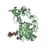
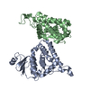
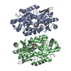
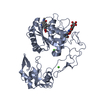
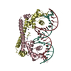
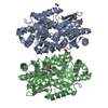
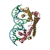
| Title | Crystal structure of VAP2 from Crotalus atrox venom (Form 2-5 crystal) | |||||||||
 Components Components | Catrocollastatin | |||||||||
 Keywords Keywords | APOPTOSIS / TOXIN / apoptotic toxin / SVMP / metalloproteinase | |||||||||
| Function / homology |  Function and homology information Function and homology informationHydrolases; Acting on peptide bonds (peptidases); Metalloendopeptidases / metalloendopeptidase activity / toxin activity / proteolysis / extracellular region / metal ion binding / plasma membrane Similarity search - Function | |||||||||
| Biological species |  Crotalus atrox (western diamondback rattlesnake) Crotalus atrox (western diamondback rattlesnake) | |||||||||
| Method |  X-RAY DIFFRACTION / SYNCHROTRON / MOLECULAR REPLACEMENT / Resolution: 2.7 Å X-RAY DIFFRACTION / SYNCHROTRON / MOLECULAR REPLACEMENT / Resolution: 2.7 Å | |||||||||
 Authors Authors | Takeda, S. / Igarashi, T. / Araki, S. | |||||||||
 Citation Citation | Journal: Febs Lett. / Year: 2007 Title: Crystal structures of catrocollastatin/VAP2B reveal a dynamic, modular architecture of ADAM/adamalysin/reprolysin family proteins Authors: Igarashi, T. / Araki, S. / Mori, H. / Takeda, S. #1: Journal: ACTA CRYSTALLOGR.,SECT.F / Year: 2006 Title: Crystallization and preliminary X-ray crystallographic analysis of two vascular apoptosis-inducing proteins (VAPs) from Crotalus atrox venom. Authors: Igarashi, T. / Oishi, Y. / Araki, S. / Mori, H. / Takeda, S. #2: Journal: Embo J. / Year: 2006Title: Crystal structures of VAP1 reveal ADAMs' MDC domain architecture and its unique C-shaped scaffold Authors: Takeda, S. / Igarashi, T. / Mori, H. / Araki, S. #3: Journal: ENDOTHELIUM / Year: 2007 Title: cDNA cloning and some additional peptide characterization of a single-chain vascular apoptosis-inducing protein, VAP2 Authors: Masuda, S. / Maeda, H. / Miao, J.Y. / Hayashi, H. / Araki, S. #4: Journal: Eur.J.Biochem. / Year: 1998 Title: Two vascular apoptosis-inducing proteins from snake venom are members of the metalloprotease/disintegrin family Authors: Masuda, S. / Hayashi, H. / Araki, S. | |||||||||
| History |
| |||||||||
| Remark 999 | SEQUENCE There is difference between the SEQRES and the sequence database. The depositors believe ...SEQUENCE There is difference between the SEQRES and the sequence database. The depositors believe it is a variant. |
- Structure visualization
| Structure viewer | Molecule: MolmilJmol/JSmol |
|---|
- Downloads & links
-Download
| PDBx/mmCIF format | 2dw2.cif.gz | 183 KB | Display | PDBx/mmCIF format |
|---|---|---|---|---|
| PDB format | pdb2dw2.ent.gz | 144.6 KB | Display | PDB format |
| PDBx/mmJSON format | 2dw2.json.gz | Tree view | PDBx/mmJSON format | |
| Others |  Other downloads Other downloads |
-Validation report
| Summary document | 2dw2_validation.pdf.gz | 1.2 MB | Display | wwPDB validaton report |
|---|---|---|---|---|
| Full document | 2dw2_full_validation.pdf.gz | 1.2 MB | Display | |
| Data in XML | 2dw2_validation.xml.gz | 34.2 KB | Display | |
| Data in CIF | 2dw2_validation.cif.gz | 47.1 KB | Display | |
| Arichive directory | https://data.pdbj.org/pub/pdb/validation_reports/dw/2dw2ftp://data.pdbj.org/pub/pdb/validation_reports/dw/2dw2 | HTTPS FTP |
-Related structure data
| Related structure data |  2dw0C  2dw1C 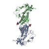 2eroS C: citing same article ( S: Starting model for refinement |
|---|---|
| Similar structure data |
-Links
 PDBj
PDBj


- Assembly
| Deposited unit | 
| ||||||||
|---|---|---|---|---|---|---|---|---|---|
| 1 | 
| ||||||||
| 2 | 
| ||||||||
| Unit cell |
|
-Components
| #1: Protein | Mass: 46912.762 Da / Num. of mol.: 2 / Fragment: residues 191-609 / Source method: isolated from a natural source Source: (natural) Crotalus atrox (western diamondback rattlesnake)References: UniProt: Q90282 #2: Polysaccharide | Source method: isolated from a genetically manipulated source #3: Chemical |   Mass: 65.409 Da / Num. of mol.: 2 / Source method: obtained synthetically / Formula: Zn Mass: 65.409 Da / Num. of mol.: 2 / Source method: obtained synthetically / Formula: Zn#4: Chemical | ChemComp-CA /   Mass: 40.078 Da / Num. of mol.: 6 / Source method: obtained synthetically / Formula: Ca Mass: 40.078 Da / Num. of mol.: 6 / Source method: obtained synthetically / Formula: Ca#5: Water | ChemComp-HOH / |  Mass: 18.015 Da / Num. of mol.: 151 / Source method: isolated from a natural source / Formula: H2O Mass: 18.015 Da / Num. of mol.: 151 / Source method: isolated from a natural source / Formula: H2OHas protein modification | Y | |
|---|
-Experimental details
-Experiment
| Experiment | Method: X-RAY DIFFRACTION / Number of used crystals: 1 |
|---|
- Sample preparation
| Crystal | Density Matthews: 2.74 Å3/Da / Density % sol: 55.13 % |
|---|---|
| Crystal grow | Temperature: 293 K / Method: vapor diffusion, sitting drop / pH: 6.5 Details: 4% n-propanol, 16.2% PEG8000, 0.18M calcium acetate, 0.09M sodium cacodylate, pH 6.5, VAPOR DIFFUSION, SITTING DROP, temperature 293K |
-Data collection
| Diffraction | Mean temperature: 90 K |
|---|---|
| Diffraction source | Source: SYNCHROTRON / Site: SPring-8  / Beamline: BL41XU / Wavelength: 1 Å / Beamline: BL41XU / Wavelength: 1 Å |
| Detector | Type: ADSC QUANTUM 315 / Detector: CCD / Date: Nov 20, 2005 |
| Radiation | Monochromator: rotated-inclined double-crystal monochromator Protocol: SINGLE WAVELENGTH / Monochromatic (M) / Laue (L): M / Scattering type: x-ray |
| Radiation wavelength | Wavelength: 1 Å / Relative weight: 1 |
| Reflection | Resolution: 2.7→50 Å / Num. all: 28047 / Num. obs: 26911 / % possible obs: 95.9 % / Observed criterion σ(F): 0 / Observed criterion σ(I): 0 / Redundancy: 3.4 % / Rmerge(I) obs: 0.085 / Net I/σ(I): 10.1 |
| Reflection shell | Resolution: 2.7→2.8 Å / Redundancy: 2.8 % / Rmerge(I) obs: 0.231 / Mean I/σ(I) obs: 5.5 / Num. unique all: 2313 / % possible all: 82.5 |
- Processing
| Software |
| |||||||||||||||||||||||||
|---|---|---|---|---|---|---|---|---|---|---|---|---|---|---|---|---|---|---|---|---|---|---|---|---|---|---|
| Refinement | Method to determine structure: MOLECULAR REPLACEMENT Starting model: 2ERO Resolution: 2.7→50 Å / Cross valid method: THROUGHOUT / σ(F): 0 / Stereochemistry target values: Engh & Huber
| |||||||||||||||||||||||||
| Refinement step | Cycle: LAST / Resolution: 2.7→50 Å
| |||||||||||||||||||||||||
| Refine LS restraints |
| |||||||||||||||||||||||||
| LS refinement shell | Resolution: 2.7→2.8 Å
|
