 Movie
Movie Controller


+ Open data

- Basic information
| Entry | Database: PDB / ID: 2dvg | |||||||||
|---|---|---|---|---|---|---|---|---|---|---|
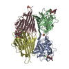
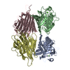
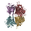
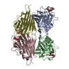
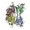
| Title | Crystal structure of peanut lectin GAL-ALPHA-1,6-GLC complex | |||||||||
 Components Components | Galactose-binding lectin | |||||||||
 Keywords Keywords | SUGAR BINDING PROTEIN / LEGUME LECTIN / AGGLUTININ / OPEN QUATERNARY STRUCTURE / CARBOHYDRATE SPECIFICITY | |||||||||
| Function / homology |  Function and homology information Function and homology information | |||||||||
| Biological species |  | |||||||||
| Method |  X-RAY DIFFRACTION / MOLECULAR REPLACEMENT / Resolution: 2.78 Å X-RAY DIFFRACTION / MOLECULAR REPLACEMENT / Resolution: 2.78 Å | |||||||||
 Authors Authors | Natchiar, S.K. / Srinivas, O. / Mitra, N. / Surolia, A. / Jayaraman, N. / Vijayan, M. | |||||||||
 Citation Citation | Journal: ACTA CRYSTALLOGR.,SECT.D / Year: 2006 Title: Structural studies on peanut lectin complexed with disaccharides involving different linkages: further insights into the structure and interactions of the lectin Authors: Natchiar, S.K. / Srinivas, O. / Mitra, N. / Surolia, A. / Jayaraman, N. / Vijayan, M. #1: Journal: Proc.Natl.Acad.Sci.USA / Year: 1994 Title: Crystal structure of peanut lectin, a protein with an unusual quaternary structure Authors: Banerjee, R. / Mande, S.C. / Ganesh, V. / Das, K. / Dhanaraj, V. / Mahanta, S.K. / Suguna, K. / Surolia, A. / Vijayan, M. #2: Journal: J.Mol.Biol. / Year: 1996Title: Conformation, protein-carbohydrate interactions and a novel subunit association in the refined structure of peanut lectin-lactose complex Authors: Banerjee, R. / Das, K. / Ravishankar, R. / Suguna, K. / Surolia, A. / Vijayan, M. #3: Journal: Curr.Sci. / Year: 1997Title: The Specificity of Peanut Agglutinin for Thomsen-Friedenreich Antigen is Mediated by Water-Bridges Authors: Ravishankar, R. / Ravindran, M. / Suguna, K. / Surolia, A. / Vijayan, M. #4: Journal: ACTA CRYSTALLOGR.,SECT.D / Year: 2004Title: Structural plasticity of peanut lectin: an X-ray analysis involving variation in pH, ligand binding and crystal structure Authors: Natchiar, S.K. / Jeyaprakash, A.A. / Ramya, T.N. / Thomas, C.J. / Suguna, K. / Surolia, A. / Vijayan, M. #5: Journal: Curr.Sci. / Year: 2006Title: Multivalency in Lectins. A Crystallographic Modellin and Light-Scattering Study Involving Peanut Lectin and a Bivalent Ligand Authors: Natchiar, S.K. / Srinivas, O. / Nivedita, M. / Sagarika, D. / Jayaraman, N. / Surolia, A. / Vijayan, M. | |||||||||
| History |
|
- Structure visualization
| Structure viewer | Molecule: MolmilJmol/JSmol |
|---|
- Downloads & links
-Download
| PDBx/mmCIF format | 2dvg.cif.gz | 197.3 KB | Display | PDBx/mmCIF format |
|---|---|---|---|---|
| PDB format | pdb2dvg.ent.gz | 157 KB | Display | PDB format |
| PDBx/mmJSON format | 2dvg.json.gz | Tree view | PDBx/mmJSON format | |
| Others |  Other downloads Other downloads |
-Validation report
| Arichive directory | https://data.pdbj.org/pub/pdb/validation_reports/dv/2dvgftp://data.pdbj.org/pub/pdb/validation_reports/dv/2dvg | HTTPS FTP |
|---|
-Related structure data
| Related structure data |  2dv9C  2dvaC  2dvbC  2dvdC  2dvfC 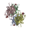 2pelS S: Starting model for refinement C: citing same article ( |
|---|---|
| Similar structure data |


-Links
 PDBj
PDBj

- Assembly
| Deposited unit | 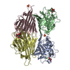
| ||||||||
|---|---|---|---|---|---|---|---|---|---|
| 1 |
| ||||||||
| Unit cell |
| ||||||||
| Details | Biological unit is a tetramer. Asymmetric unit contains one biological unit. |
-Components
-Protein , 1 types, 4 molecules ABCD
| #1: Protein | Mass: 25208.955 Da / Num. of mol.: 4 / Fragment: residues 1-236 / Source method: isolated from a natural source / Source: (natural) |
|---|
-Sugars , 2 types, 4 molecules 
| #2: Polysaccharide | alpha-D-galactopyranose-(1-6)-beta-D-glucopyranose Source method: isolated from a genetically manipulated source |
|---|---|
| #3: Sugar |  Type: D-saccharide, alpha linking / Mass: 180.156 Da / Num. of mol.: 3 Type: D-saccharide, alpha linking / Mass: 180.156 Da / Num. of mol.: 3Source method: isolated from a genetically manipulated source Formula: C6H12O6 |
-Non-polymers , 4 types, 476 molecules 


| #4: Chemical | ChemComp-CA /  Mass: 40.078 Da / Num. of mol.: 4 / Source method: obtained synthetically / Formula: Ca Mass: 40.078 Da / Num. of mol.: 4 / Source method: obtained synthetically / Formula: Ca#5: Chemical | ChemComp-MN /  Mass: 54.938 Da / Num. of mol.: 4 / Source method: obtained synthetically / Formula: Mn Mass: 54.938 Da / Num. of mol.: 4 / Source method: obtained synthetically / Formula: Mn#6: Chemical |  Mass: 96.063 Da / Num. of mol.: 2 / Source method: obtained synthetically / Formula: SO4 Mass: 96.063 Da / Num. of mol.: 2 / Source method: obtained synthetically / Formula: SO4#7: Water | ChemComp-HOH / | Mass: 18.015 Da / Num. of mol.: 466 / Source method: isolated from a natural source / Formula: H2O |
|---|
-Experimental details
-Experiment
| Experiment | Method: X-RAY DIFFRACTION / Number of used crystals: 1 |
|---|
- Sample preparation
| Crystal | Density Matthews: 2.7 Å3/Da / Density % sol: 54.2 % |
|---|---|
| Crystal grow | Temperature: 288 K / Method: vapor diffusion, hanging drop / pH: 6.5 Details: 7.5% PEG 8000, 0.025M Sodium cadodylate, 0.5M ammonium sulfate, pH 6.5, VAPOR DIFFUSION, HANGING DROP, temperature 288K |
-Data collection
| Diffraction | Mean temperature: 100 K |
|---|---|
| Diffraction source | Source: ROTATING ANODE / Type: RIGAKU RU200 / Wavelength: 1.5418 Å |
| Detector | Type: MAR scanner 345 mm plate / Detector: IMAGE PLATE |
| Radiation | Monochromator: Osmic mirror / Protocol: SINGLE WAVELENGTH / Monochromatic (M) / Laue (L): M / Scattering type: x-ray |
| Radiation wavelength | Wavelength: 1.5418 Å / Relative weight: 1 |
| Reflection | Resolution: 2.78→20 Å / Num. obs: 29220 / % possible obs: 95.2 % / Redundancy: 5.3 % / Biso Wilson estimate: 55.8 Å2 / Rmerge(I) obs: 0.167 / Rsym value: 0.185 / Net I/σ(I): 8.8 |
| Reflection shell | Resolution: 2.74→2.78 Å / Redundancy: 5.3 % / Rmerge(I) obs: 0.498 / Mean I/σ(I) obs: 4.5 / Num. unique all: 2865 / Rsym value: 0.556 / % possible all: 95.1 |
- Processing
| Software |
| ||||||||||||||||||||||||||||||||||||
|---|---|---|---|---|---|---|---|---|---|---|---|---|---|---|---|---|---|---|---|---|---|---|---|---|---|---|---|---|---|---|---|---|---|---|---|---|---|
| Refinement | Method to determine structure: MOLECULAR REPLACEMENT Starting model: 2PEL Resolution: 2.78→19.93 Å / Rfactor Rfree error: 0.007 / Data cutoff high absF: 2049486.95 / Data cutoff low absF: 0 / Isotropic thermal model: RESTRAINED / Cross valid method: THROUGHOUT / σ(F): 0 / Details: MLF TARGET
| ||||||||||||||||||||||||||||||||||||
| Solvent computation | Solvent model: FLAT MODEL / Bsol: 46.016 Å2 / ksol: 0.324288 e/Å3 | ||||||||||||||||||||||||||||||||||||
| Displacement parameters | Biso mean: 32.7 Å2
| ||||||||||||||||||||||||||||||||||||
| Refine analyze |
| ||||||||||||||||||||||||||||||||||||
| Refinement step | Cycle: LAST / Resolution: 2.78→19.93 Å
| ||||||||||||||||||||||||||||||||||||
| Refine LS restraints |
| ||||||||||||||||||||||||||||||||||||
| LS refinement shell | Resolution: 2.78→2.95 Å / Rfactor Rfree error: 0.022 / Total num. of bins used: 6
|
