 Movie
Movie Controller


+ Open data

- Basic information
| Entry | Database: PDB / ID: 1cq9 | ||||||
|---|---|---|---|---|---|---|---|
| Title | PEANUT LECTIN-TRICLINIC FORM | ||||||
 Components Components | PROTEIN (PEANUT LECTIN) | ||||||
 Keywords Keywords | LECTIN / LEGUME LECTIN / OPEN QUATERNARY STRUCTURE / TRICLINIC FORM / ACIDIC PH | ||||||
| Function / homology |  Function and homology information Function and homology information | ||||||
| Biological species |  | ||||||
| Method |  X-RAY DIFFRACTION / MOLECULAR REPLACEMENT / Resolution: 3.5 Å X-RAY DIFFRACTION / MOLECULAR REPLACEMENT / Resolution: 3.5 Å | ||||||
 Authors Authors | Ravishankar, R. / Suguna, K. / Surolia, A. / Vijayan, M. | ||||||
 Citation Citation | Journal: Proteins / Year: 2001 Title: Crystal structures of the peanut lectin-lactose complex at acidic pH: retention of unusual quaternary structure, empty and carbohydrate bound combining sites, molecular mimicry and crystal ...Title: Crystal structures of the peanut lectin-lactose complex at acidic pH: retention of unusual quaternary structure, empty and carbohydrate bound combining sites, molecular mimicry and crystal packing directed by interactions at the combining site. Authors: Ravishankar, R. / Thomas, C.J. / Suguna, K. / Surolia, A. / Vijayan, M. #1: Journal: Acta Crystallogr.,Sect.D / Year: 1999Title: Structures of the Complexes of Peanut Lectin with Methyl-Beta-Galactose and N- Acetyllactosamine and a Comparative Study of Carbohydrate Binding in Gal/ Galnac-Specific Legume Lectins Authors: Ravishankar, R. / Suguna, K. / Surolia, A. / Vijayan, M. #2: Journal: Curr.Sci. / Year: 1997Title: The Specificity of Peanut Agglutinin for Thomsen-Friedenreich Antigen is Mediated by Water-Bridges Authors: Ravishankar, R. / Ravindran, M. / Suguna, K. / Surolia, A. / Vijayan, M. #3: Journal: J.Mol.Biol. / Year: 1996Title: Conformation, Protein-Carbohydrate Interactions and a Novel Subunit Association in the Refined Structure of Peanut Lectin-Lactose Complex Authors: Banerjee, R. / Das, K. / Ravishankar, R. / Suguna, K. / Surolia, A. / Vijayan, M. #4: Journal: Proc.Natl.Acad.Sci.USA / Year: 1994Title: Crystal Structure of Peanut Lectin, a Protein with an Unusual Quaternary Structure Authors: Banerjee, R. / Mande, S.C. / Ganesh, V. / Das, K. / Dhanaraj, V. / Mahanta, S.K. / Suguna, K. / Surolia, A. / Vijayan, M. | ||||||
| History |
|
- Structure visualization
| Structure viewer | Molecule: MolmilJmol/JSmol |
|---|
- Downloads & links
-Download
| PDBx/mmCIF format | 1cq9.cif.gz | 179.1 KB | Display | PDBx/mmCIF format |
|---|---|---|---|---|
| PDB format | pdb1cq9.ent.gz | 142.7 KB | Display | PDB format |
| PDBx/mmJSON format | 1cq9.json.gz | Tree view | PDBx/mmJSON format | |
| Others |  Other downloads Other downloads |
-Validation report
| Arichive directory | https://data.pdbj.org/pub/pdb/validation_reports/cq/1cq9ftp://data.pdbj.org/pub/pdb/validation_reports/cq/1cq9 | HTTPS FTP |
|---|
-Related structure data
| Related structure data |  1cr7C 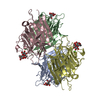 2pelS S: Starting model for refinement C: citing same article ( |
|---|---|
| Similar structure data |


-Links
 PDBj
PDBj

- Assembly
| Deposited unit | 
| ||||||||
|---|---|---|---|---|---|---|---|---|---|
| 1 |
| ||||||||
| Unit cell |
|
-Components
| #1: Protein | Mass: 25208.955 Da / Num. of mol.: 4 / Source method: isolated from a natural source / Source: (natural) #2: Chemical | ChemComp-CA /   Mass: 40.078 Da / Num. of mol.: 4 / Source method: obtained synthetically / Formula: Ca Mass: 40.078 Da / Num. of mol.: 4 / Source method: obtained synthetically / Formula: Ca#3: Chemical | ChemComp-MN /   Mass: 54.938 Da / Num. of mol.: 4 / Source method: obtained synthetically / Formula: Mn Mass: 54.938 Da / Num. of mol.: 4 / Source method: obtained synthetically / Formula: Mn |
|---|
-Experimental details
-Experiment
| Experiment | Method: X-RAY DIFFRACTION / Number of used crystals: 1 |
|---|
- Sample preparation
| Crystal | Density Matthews: 2.84 Å3/Da / Density % sol: 56.72 % | ||||||||||||||||||||||||||||||||||||||||||
|---|---|---|---|---|---|---|---|---|---|---|---|---|---|---|---|---|---|---|---|---|---|---|---|---|---|---|---|---|---|---|---|---|---|---|---|---|---|---|---|---|---|---|---|
| Crystal grow | pH: 4.6 / Details: pH 4.6 | ||||||||||||||||||||||||||||||||||||||||||
| Crystal grow | *PLUS Temperature: 20 ℃ / pH: 7.4 / Method: microdialysis / Details: Salunke, D.M., (1983) FEBS Lett., 156, 127. | ||||||||||||||||||||||||||||||||||||||||||
| Components of the solutions | *PLUS
|
-Data collection
| Diffraction | Mean temperature: 293 K |
|---|---|
| Diffraction source | Source: ROTATING ANODE / Type: RIGAKU RU200 / Wavelength: 1.5418 |
| Detector | Type: MARRESEARCH / Detector: IMAGE PLATE |
| Radiation | Protocol: SINGLE WAVELENGTH / Monochromatic (M) / Laue (L): M / Scattering type: x-ray |
| Radiation wavelength | Wavelength: 1.5418 Å / Relative weight: 1 |
| Reflection | Resolution: 3→28.7 Å / Num. obs: 18342 / % possible obs: 82.32 % / Redundancy: 1.39 % / Rmerge(I) obs: 0.1572 / Net I/σ(I): 2.53 |
| Reflection | *PLUS Highest resolution: 3.5 Å / Num. obs: 11935 / % possible obs: 84.3 % / Num. measured all: 16538 / Rmerge(I) obs: 0.157 |
| Reflection shell | *PLUS % possible obs: 81.4 % / Rmerge(I) obs: 0.301 |
- Processing
| Software |
| ||||||||||||||||||||||||||||||||||||||||||||||||||||||||||||
|---|---|---|---|---|---|---|---|---|---|---|---|---|---|---|---|---|---|---|---|---|---|---|---|---|---|---|---|---|---|---|---|---|---|---|---|---|---|---|---|---|---|---|---|---|---|---|---|---|---|---|---|---|---|---|---|---|---|---|---|---|---|
| Refinement | Method to determine structure: MOLECULAR REPLACEMENT Starting model: 2PEL WITHOUT SUGAR AND WATER Resolution: 3.5→10 Å / Rfactor Rfree error: 0.012 / Data cutoff high absF: 10000000 / Data cutoff low absF: 0 / Cross valid method: THROUGHOUT / σ(F): 0 Details: THERE IS A TETRAMER IN THE UNIT CELL WITHOUT 222 OR FOUR-FOLD SYMMETRY.
| ||||||||||||||||||||||||||||||||||||||||||||||||||||||||||||
| Displacement parameters | Biso mean: 20.1 Å2
| ||||||||||||||||||||||||||||||||||||||||||||||||||||||||||||
| Refine analyze |
| ||||||||||||||||||||||||||||||||||||||||||||||||||||||||||||
| Refinement step | Cycle: LAST / Resolution: 3.5→10 Å
| ||||||||||||||||||||||||||||||||||||||||||||||||||||||||||||
| Refine LS restraints |
| ||||||||||||||||||||||||||||||||||||||||||||||||||||||||||||
| Refine LS restraints NCS | NCS model details: RESTRAIN | ||||||||||||||||||||||||||||||||||||||||||||||||||||||||||||
| LS refinement shell | Resolution: 3.5→3.71 Å / Rfactor Rfree error: 0.039 / Total num. of bins used: 6
| ||||||||||||||||||||||||||||||||||||||||||||||||||||||||||||
| Xplor file |
| ||||||||||||||||||||||||||||||||||||||||||||||||||||||||||||
| Refinement | *PLUS Highest resolution: 3.5 Å / Lowest resolution: 10 Å / Rfactor obs: 0.217 / Rfactor Rfree: 0.278 / Rfactor Rwork: 0.217 | ||||||||||||||||||||||||||||||||||||||||||||||||||||||||||||
| Solvent computation | *PLUS | ||||||||||||||||||||||||||||||||||||||||||||||||||||||||||||
| Displacement parameters | *PLUS | ||||||||||||||||||||||||||||||||||||||||||||||||||||||||||||
| Refine LS restraints | *PLUS
| ||||||||||||||||||||||||||||||||||||||||||||||||||||||||||||
| LS refinement shell | *PLUS Rfactor Rfree: 0.276 / Rfactor Rwork: 0.237 |
