 Movie
Movie Controller


+ Open data

- Basic information
| Entry | Database: PDB / ID: 2dtr | |||||||||
|---|---|---|---|---|---|---|---|---|---|---|
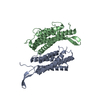
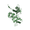
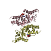
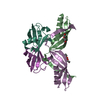
| Title | STRUCTURE OF DIPHTHERIA TOXIN REPRESSOR | |||||||||
 Components Components | DIPHTHERIA TOXIN REPRESSOR | |||||||||
 Keywords Keywords | REPRESSOR / TRANSCRIPTION REGULATION / DNA-BINDING / IRON | |||||||||
| Function / homology |  Function and homology information Function and homology informationtransition metal ion binding / SH3 domain binding / protein dimerization activity / DNA-binding transcription factor activity / negative regulation of DNA-templated transcription / DNA binding / identical protein binding / cytoplasm Similarity search - Function | |||||||||
| Biological species |  Corynebacterium diphtheriae (bacteria) Corynebacterium diphtheriae (bacteria) | |||||||||
| Method |  X-RAY DIFFRACTION / Resolution: 1.9 Å X-RAY DIFFRACTION / Resolution: 1.9 Å | |||||||||
 Authors Authors | Qiu, X. / Pohl, E. / Hol, W.G. | |||||||||
 Citation Citation | Journal: Biochemistry / Year: 1996 Title: High-resolution structure of the diphtheria toxin repressor complexed with cobalt and manganese reveals an SH3-like third domain and suggests a possible role of phosphate as co-corepressor. Authors: Qiu, X. / Pohl, E. / Holmes, R.K. / Hol, W.G. #1: Journal: Proc.Natl.Acad.Sci.USA / Year: 1992Title: Purification and Characterization of the Diphtheria Toxin Repressor Authors: Schmitt, M.P. / Twiddy, E.M. / Holmes, R.K. | |||||||||
| History |
|
- Structure visualization
| Structure viewer | Molecule: MolmilJmol/JSmol |
|---|
- Downloads & links
-Download
| PDBx/mmCIF format | 2dtr.cif.gz | 49.6 KB | Display | PDBx/mmCIF format |
|---|---|---|---|---|
| PDB format | pdb2dtr.ent.gz | 31.4 KB | Display | PDB format |
| PDBx/mmJSON format | 2dtr.json.gz | Tree view | PDBx/mmJSON format | |
| Others |  Other downloads Other downloads |
-Validation report
| Summary document | 2dtr_validation.pdf.gz | 379.3 KB | Display | wwPDB validaton report |
|---|---|---|---|---|
| Full document | 2dtr_full_validation.pdf.gz | 379.7 KB | Display | |
| Data in XML | 2dtr_validation.xml.gz | 4.3 KB | Display | |
| Data in CIF | 2dtr_validation.cif.gz | 7.5 KB | Display | |
| Arichive directory | https://data.pdbj.org/pub/pdb/validation_reports/dt/2dtrftp://data.pdbj.org/pub/pdb/validation_reports/dt/2dtr | HTTPS FTP |
-Related structure data
| Similar structure data |
|---|


-Links
 PDBj
PDBj
- Assembly
| Deposited unit | 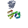
| ||||||||
|---|---|---|---|---|---|---|---|---|---|
| 1 | 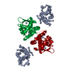
| ||||||||
| Unit cell |
|
-Components
| #1: Protein | Mass: 25349.795 Da / Num. of mol.: 1 Source method: isolated from a genetically manipulated source Source: (gene. exp.) Corynebacterium diphtheriae (bacteria) / Strain: C7(-) / Gene: T / Plasmid: PMS298 / Gene (production host): T / Production host: |
|---|---|
| #2: Chemical | ChemComp-SO4 /   Mass: 96.063 Da / Num. of mol.: 1 / Source method: obtained synthetically / Formula: SO4 Mass: 96.063 Da / Num. of mol.: 1 / Source method: obtained synthetically / Formula: SO4 |
| #3: Chemical | ChemComp-CO /   Mass: 58.933 Da / Num. of mol.: 1 / Source method: obtained synthetically / Formula: Co Mass: 58.933 Da / Num. of mol.: 1 / Source method: obtained synthetically / Formula: Co |
| #4: Water | ChemComp-HOH /  Mass: 18.015 Da / Num. of mol.: 148 / Source method: isolated from a natural source / Formula: H2O Mass: 18.015 Da / Num. of mol.: 148 / Source method: isolated from a natural source / Formula: H2O |
-Experimental details
-Experiment
| Experiment | Method: X-RAY DIFFRACTION |
|---|
- Sample preparation
| Crystal | Density Matthews: 2.54 Å3/Da / Density % sol: 57 % | ||||||||||||||||||||||||||||||||||||||||||||||||
|---|---|---|---|---|---|---|---|---|---|---|---|---|---|---|---|---|---|---|---|---|---|---|---|---|---|---|---|---|---|---|---|---|---|---|---|---|---|---|---|---|---|---|---|---|---|---|---|---|---|
| Crystal grow | *PLUS Method: vapor diffusion, hanging drop | ||||||||||||||||||||||||||||||||||||||||||||||||
| Components of the solutions | *PLUS
|
-Data collection
| Diffraction source | Wavelength: 1.5418 |
|---|---|
| Detector | Type: RIGAKU RAXIS / Detector: IMAGE PLATE / Date: Oct 1, 1995 |
| Radiation | Monochromatic (M) / Laue (L): M / Scattering type: x-ray |
| Radiation wavelength | Wavelength: 1.5418 Å / Relative weight: 1 |
| Reflection | Num. obs: 14788 / % possible obs: 78 % / Redundancy: 3 % / Rmerge(I) obs: 0.045 |
| Reflection | *PLUS Highest resolution: 1.9 Å / Lowest resolution: 50 Å / Num. measured all: 41868 / Rmerge(I) obs: 0.045 |
| Reflection shell | *PLUS Highest resolution: 2 Å / Lowest resolution: 2.25 Å / % possible obs: 52 % |
- Processing
| Software |
| ||||||||||||||||||||||||||||||||||||||||||||||||||||||||||||
|---|---|---|---|---|---|---|---|---|---|---|---|---|---|---|---|---|---|---|---|---|---|---|---|---|---|---|---|---|---|---|---|---|---|---|---|---|---|---|---|---|---|---|---|---|---|---|---|---|---|---|---|---|---|---|---|---|---|---|---|---|---|
| Refinement | Resolution: 1.9→7 Å / σ(F): 1 Details: 2FO-FC ELECTRON DENSITY IS MISSING FOR RESIDUES 1 - 3, 141 - 147 AND 199 - 200. THESE RESIDUES ARE ASSUMED TO BE DISORDERED. THE EXPERIMENTAL ELECTRON DENSITY IS WEAK OR MISSING FOR RESIDUES 148 - 226.
| ||||||||||||||||||||||||||||||||||||||||||||||||||||||||||||
| Refinement step | Cycle: LAST / Resolution: 1.9→7 Å
| ||||||||||||||||||||||||||||||||||||||||||||||||||||||||||||
| Refine LS restraints |
| ||||||||||||||||||||||||||||||||||||||||||||||||||||||||||||
| Software | *PLUS Name: X-PLOR / Classification: refinement | ||||||||||||||||||||||||||||||||||||||||||||||||||||||||||||
| Refinement | *PLUS Rfactor obs: 0.0171 / Lowest resolution: 7 Å | ||||||||||||||||||||||||||||||||||||||||||||||||||||||||||||
| Solvent computation | *PLUS | ||||||||||||||||||||||||||||||||||||||||||||||||||||||||||||
| Displacement parameters | *PLUS | ||||||||||||||||||||||||||||||||||||||||||||||||||||||||||||
| Refine LS restraints | *PLUS
|
