[English] 日本語
Yorodumi
- PDB-2dt3: Crystal structure of the complex formed between goat signalling p... -

+
Open data


ID or keywords:

Loading...

-
Basic information

Entry
Database: PDB / ID: 2dt3
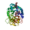
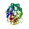
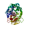
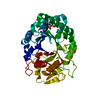
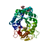
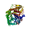
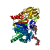
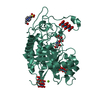
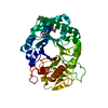
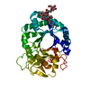
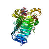
TitleCrystal structure of the complex formed between goat signalling protein and the hexasaccharide at 2.28 A resolution
ComponentsChitinase-3-like protein 1
KeywordsSIGNALING PROTEIN / TIM barrel / Signalling / Involution / Mammary gland
Function / homology
Function and homology information


response to interleukin-6 / activation of NF-kappaB-inducing kinase activity / chitin binding / response to tumor necrosis factor / response to mechanical stimulus / response to interleukin-1 / positive regulation of peptidyl-threonine phosphorylation / positive regulation of interleukin-8 production / lung development / positive regulation of angiogenesis ...response to interleukin-6 / activation of NF-kappaB-inducing kinase activity / chitin binding / response to tumor necrosis factor / response to mechanical stimulus / response to interleukin-1 / positive regulation of peptidyl-threonine phosphorylation / positive regulation of interleukin-8 production / lung development / positive regulation of angiogenesis / cellular response to tumor necrosis factor / carbohydrate binding / positive regulation of ERK1 and ERK2 cascade / positive regulation of phosphatidylinositol 3-kinase/protein kinase B signal transduction / carbohydrate metabolic process / inflammatory response / apoptotic process / perinuclear region of cytoplasm / endoplasmic reticulum / extracellular space / cytoplasm
Similarity search - Function
Chitinase A; domain 3 - #10 / Chitinase insertion domain superfamily / Chitinase II / Glyco_18 / Glycosyl hydrolases family 18 (GH18) domain profile. / Glycoside hydrolase family 18, catalytic domain / Glycosyl hydrolases family 18 / Chitinase A; domain 3 / Glycosidases / Glycoside hydrolase superfamily ...Chitinase A; domain 3 - #10 / Chitinase insertion domain superfamily / Chitinase II / Glyco_18 / Glycosyl hydrolases family 18 (GH18) domain profile. / Glycoside hydrolase family 18, catalytic domain / Glycosyl hydrolases family 18 / Chitinase A; domain 3 / Glycosidases / Glycoside hydrolase superfamily / TIM Barrel / Roll / Alpha-Beta Barrel / Alpha Beta
Similarity search - Domain/homology
Chitinase-3-like protein 1
Similarity search - Component
Biological speciesCapra hircus (goat)
MethodX-RAY DIFFRACTION / MOLECULAR REPLACEMENT / Resolution: 2.28 Å
AuthorsKumar, J. / Ethayathulla, A.S. / Srivastava, D.B. / Singh, N. / Sharma, S. / Singh, T.P.
CitationJournal: ACTA CRYSTALLOGR.,SECT.D / Year: 2007
Title: Carbohydrate-binding properties of goat secretory glycoprotein (SPG-40) and its functional implications: structures of the native glycoprotein and its four complexes with chitin-like oligosaccharides
Authors: Kumar, J. / Ethayathulla, A.S. / Srivastava, D.B. / Singh, N. / Sharma, S. / Kaur, P. / Srinivasan, A. / Singh, T.P.
History
DepositionJul 9, 2006Deposition site: PDBJ / Processing site: PDBJ
SupersessionAug 1, 2006ID: 2B2Z
Revision 1.0Aug 1, 2006Provider: repository / Type: Initial release
Revision 1.1Apr 30, 2008Group: Version format compliance
Revision 1.2Jul 13, 2011Group: Non-polymer description / Version format compliance
Revision 2.0Jul 29, 2020Group: Advisory / Atomic model ...Advisory / Atomic model / Data collection / Derived calculations / Structure summary
Category: atom_site / chem_comp ...atom_site / chem_comp / database_PDB_caveat / entity / pdbx_branch_scheme / pdbx_chem_comp_identifier / pdbx_entity_branch / pdbx_entity_branch_descriptor / pdbx_entity_branch_link / pdbx_entity_branch_list / pdbx_entity_nonpoly / pdbx_nonpoly_scheme / pdbx_struct_assembly_gen / pdbx_validate_chiral / struct_asym / struct_conn / struct_site / struct_site_gen
Item: _atom_site.auth_asym_id / _atom_site.auth_seq_id ..._atom_site.auth_asym_id / _atom_site.auth_seq_id / _atom_site.label_asym_id / _atom_site.label_entity_id / _chem_comp.name / _chem_comp.type / _entity.formula_weight / _entity.pdbx_description / _entity.pdbx_number_of_molecules / _entity.type / _pdbx_struct_assembly_gen.asym_id_list / _pdbx_validate_chiral.auth_asym_id / _pdbx_validate_chiral.auth_seq_id / _struct_conn.pdbx_leaving_atom_flag / _struct_conn.pdbx_role / _struct_conn.ptnr1_auth_asym_id / _struct_conn.ptnr1_auth_seq_id / _struct_conn.ptnr1_label_asym_id / _struct_conn.ptnr2_auth_asym_id / _struct_conn.ptnr2_auth_seq_id / _struct_conn.ptnr2_label_asym_id
Description: Carbohydrate remediation / Provider: repository / Type: Remediation
Revision 2.1Oct 25, 2023Group: Data collection / Database references ...Data collection / Database references / Refinement description / Structure summary
Category: chem_comp / chem_comp_atom ...chem_comp / chem_comp_atom / chem_comp_bond / database_2 / pdbx_initial_refinement_model
Item: _chem_comp.pdbx_synonyms / _database_2.pdbx_DOI / _database_2.pdbx_database_accession

-
Structure visualization

Structure viewerMolecule:
MolmilJmol/JSmol

Downloads & links

-
Assembly

Deposited unit
A: Chitinase-3-like protein 1
hetero molecules


Theoretical massNumber of molelcules
Total (without water)42,8763
Polymers40,7281
Non-polymers2,1482
Water3,279182
1


  • Idetical with deposited unit
  • defined by author
TypeNameSymmetry operationNumber
identity operation1_555x,y,z1
Unit cell
Length a, b, c (Å)62.300, 66.420, 106.620
Angle α, β, γ (deg.)90.00, 90.00, 90.00
Int Tables number19
Space group name H-MP212121

-
Components

#1: Protein Chitinase-3-like protein 1 / SPG-40 / Mammary gland protein MGP-40 / BP40


Mass: 40728.090 Da / Num. of mol.: 1 / Source method: isolated from a natural source / Source: (natural) Capra hircus (goat) / Secretion: MILK MAMMARY GLAND / References: UniProt: Q8SPQ0
#2: Polysaccharide alpha-D-mannopyranose-(1-4)-alpha-D-mannopyranose-(1-4)-alpha-D-mannopyranose-(1-4)-2-acetamido-2- ...alpha-D-mannopyranose-(1-4)-alpha-D-mannopyranose-(1-4)-alpha-D-mannopyranose-(1-4)-2-acetamido-2-deoxy-beta-D-glucopyranose-(1-4)-2-acetamido-2-deoxy-beta-D-glucopyranose


Type: oligosaccharide / Mass: 910.823 Da / Num. of mol.: 1
Source method: isolated from a genetically manipulated source
DescriptorTypeProgram
DManpa1-4DManpa1-4DManpa1-4DGlcpNAcb1-4DGlcpNAcb1-Glycam Condensed SequenceGMML 1.0
WURCS=2.0/2,5,4/[a2122h-1b_1-5_2*NCC/3=O][a1122h-1a_1-5]/1-1-2-2-2/a4-b1_b4-c1_c4-d1_d4-e1WURCSPDB2Glycan 1.1.0
[]{[(4+1)][b-D-GlcpNAc]{[(4+1)][a-D-GlcpNAc]{[(4+1)][b-D-Manp]{[(4+1)][a-D-Manp]{[(4+1)][b-D-Manp]{}}}}}}LINUCSPDB-CARE
#3: Polysaccharide 2-acetamido-2-deoxy-beta-D-glucopyranose-(1-4)-2-acetamido-2-deoxy-beta-D-glucopyranose-(1-4)-2- ...2-acetamido-2-deoxy-beta-D-glucopyranose-(1-4)-2-acetamido-2-deoxy-beta-D-glucopyranose-(1-4)-2-acetamido-2-deoxy-beta-D-glucopyranose-(1-4)-2-acetamido-2-deoxy-beta-D-glucopyranose-(1-4)-2-acetamido-2-deoxy-beta-D-glucopyranose-(1-4)-2-acetamido-2-deoxy-beta-D-glucopyranose


Type: oligosaccharide / Mass: 1237.172 Da / Num. of mol.: 1
Source method: isolated from a genetically manipulated source
DescriptorTypeProgram
DGlcpNAcb1-4DGlcpNAcb1-4DGlcpNAcb1-4DGlcpNAcb1-4DGlcpNAcb1-4DGlcpNAcb1-ROHGlycam Condensed SequenceGMML 1.0
WURCS=2.0/1,6,5/[a2122h-1b_1-5_2*NCC/3=O]/1-1-1-1-1-1/a4-b1_b4-c1_c4-d1_d4-e1_e4-f1WURCSPDB2Glycan 1.1.0
[][b-D-GlcpNAc]{[(4+1)][b-D-GlcpNAc]{[(4+1)][a-D-GlcpNAc]{[(4+1)][b-D-GlcpNAc]{[(4+1)][a-D-GlcpNAc]{[(4+1)][b-D-GlcpNAc]{}}}}}}LINUCSPDB-CARE
#4: Water ChemComp-HOH / water / Water


Mass: 18.015 Da / Num. of mol.: 182 / Source method: isolated from a natural source / Formula: H2O

-
Experimental details

-
Experiment

ExperimentMethod: X-RAY DIFFRACTION / Number of used crystals: 1

-
Sample preparation

CrystalDensity Matthews: 2.71 Å3/Da / Density % sol: 54.56 %
Crystal growTemperature: 298 K / Method: vapor diffusion, hanging drop / pH: 7.8
Details: Precipitant 19% Ethanol, pH 7.8, VAPOR DIFFUSION, HANGING DROP, temperature 298K

-
Data collection

DiffractionMean temperature: 298 K
Diffraction sourceSource: ROTATING ANODE / Type: RIGAKU RU300 / Wavelength: 1.5418 Å
DetectorType: MARRESEARCH / Detector: IMAGE PLATE / Date: May 10, 2005 / Details: MIRROR
RadiationMonochromator: GRAPHITE / Protocol: SINGLE WAVELENGTH / Monochromatic (M) / Laue (L): M / Scattering type: x-ray
Radiation wavelengthWavelength: 1.5418 Å / Relative weight: 1
ReflectionResolution: 2.28→56 Å / Num. obs: 21698 / % possible obs: 99.5 % / Observed criterion σ(F): 0 / Observed criterion σ(I): 0 / Biso Wilson estimate: 24.3 Å2 / Rsym value: 0.1 / Net I/σ(I): 8
Reflection shellResolution: 2.28→2.32 Å / Mean I/σ(I) obs: 2 / Rsym value: 0.49 / % possible all: 96.5

-
Processing

Software
NameVersionClassification
CNS1.1refinement
AUTOMARdata reduction
SCALEPACKdata scaling
AMoREphasing
RefinementMethod to determine structure: MOLECULAR REPLACEMENT
Starting model: 1LJY
Resolution: 2.28→56 Å / Rfactor Rfree error: 0.007 / Data cutoff high absF: 1524063.48 / Data cutoff low absF: 0 / Isotropic thermal model: RESTRAINED / Cross valid method: THROUGHOUT / σ(F): 0 / σ(I): 0 / Stereochemistry target values: Engh & Huber
RfactorNum. reflection% reflectionSelection details
Rfree0.215 1000 4.8 %RANDOM
Rwork0.196 ---
all0.197 21698 --
obs0.196 20698 99.1 %-
Solvent computationSolvent model: FLAT MODEL / Bsol: 36.5343 Å2 / ksol: 0.278407 e/Å3
Displacement parametersBiso mean: 35.8 Å2
Baniso -1Baniso -2Baniso -3
1--0.1 Å20 Å20 Å2
2---1.72 Å20 Å2
3---1.83 Å2
Refine analyze
FreeObs
Luzzati coordinate error0.28 Å0.26 Å
Luzzati d res low-5 Å
Luzzati sigma a0.33 Å0.29 Å
Refinement stepCycle: LAST / Resolution: 2.28→56 Å
ProteinNucleic acidLigandSolventTotal
Num. atoms2877 0 146 182 3205
Refine LS restraints
Refine-IDTypeDev idealDev ideal target
X-RAY DIFFRACTIONc_bond_d0.007
X-RAY DIFFRACTIONc_angle_deg1.5
X-RAY DIFFRACTIONc_dihedral_angle_d24.4
X-RAY DIFFRACTIONc_improper_angle_d0.89
X-RAY DIFFRACTIONc_mcbond_it1.531.5
X-RAY DIFFRACTIONc_mcangle_it2.662
X-RAY DIFFRACTIONc_scbond_it2.812
X-RAY DIFFRACTIONc_scangle_it42.5
LS refinement shellResolution: 2.27→2.41 Å / Rfactor Rfree error: 0.025 / Total num. of bins used: 6
RfactorNum. reflection% reflection
Rfree0.303 151 4.7 %
Rwork0.275 3030 -
obs--92.7 %
Xplor file
Refine-IDSerial noParam fileTopol file
X-RAY DIFFRACTION1protein_rep.paramprotein.top
X-RAY DIFFRACTION2ion.paramwater.top
X-RAY DIFFRACTION3water.paramion.top
X-RAY DIFFRACTION4carbohydrate.paramcarbohydrate.top
X-RAY DIFFRACTION5cis_peptide.paramcis_peptide.top

+
About Yorodumi

-
News

-
Feb 9, 2022. New format data for meta-information of EMDB entries

New format data for meta-information of EMDB entries

  • Version 3 of the EMDB header file is now the official format.
  • The previous official version 1.9 will be removed from the archive.

Related info.:EMDB header

External links:wwPDB to switch to version 3 of the EMDB data model

-
Aug 12, 2020. Covid-19 info

Covid-19 info

URL: https://pdbj.org/emnavi/covid19.php

New page: Covid-19 featured information page in EM Navigator.

Related info.:Covid-19 info / Mar 5, 2020. Novel coronavirus structure data

+
Mar 5, 2020. Novel coronavirus structure data

Novel coronavirus structure data

Related info.:Yorodumi Speices / Aug 12, 2020. Covid-19 info

External links:COVID-19 featured content - PDBj / Molecule of the Month (242):Coronavirus Proteases

+
Jan 31, 2019. EMDB accession codes are about to change! (news from PDBe EMDB page)

EMDB accession codes are about to change! (news from PDBe EMDB page)

  • The allocation of 4 digits for EMDB accession codes will soon come to an end. Whilst these codes will remain in use, new EMDB accession codes will include an additional digit and will expand incrementally as the available range of codes is exhausted. The current 4-digit format prefixed with “EMD-” (i.e. EMD-XXXX) will advance to a 5-digit format (i.e. EMD-XXXXX), and so on. It is currently estimated that the 4-digit codes will be depleted around Spring 2019, at which point the 5-digit format will come into force.
  • The EM Navigator/Yorodumi systems omit the EMD- prefix.

Related info.:Q: What is EMD? / ID/Accession-code notation in Yorodumi/EM Navigator

External links:EMDB Accession Codes are Changing Soon! / Contact to PDBj

+
Jul 12, 2017. Major update of PDB

Major update of PDB

  • wwPDB released updated PDB data conforming to the new PDBx/mmCIF dictionary.
  • This is a major update changing the version number from 4 to 5, and with Remediation, in which all the entries are updated.
  • In this update, many items about electron microscopy experimental information are reorganized (e.g. em_software).
  • Now, EM Navigator and Yorodumi are based on the updated data.

External links:wwPDB Remediation / Enriched Model Files Conforming to OneDep Data Standards Now Available in the PDB FTP Archive

-
Yorodumi

Thousand views of thousand structures

  • Yorodumi is a browser for structure data from EMDB, PDB, SASBDB, etc.
  • This page is also the successor to EM Navigator detail page, and also detail information page/front-end page for Omokage search.
  • The word "yorodu" (or yorozu) is an old Japanese word meaning "ten thousand". "mi" (miru) is to see.

Related info.:EMDB / PDB / SASBDB / Comparison of 3 databanks / Yorodumi Search / Aug 31, 2016. New EM Navigator & Yorodumi / Yorodumi Papers / Jmol/JSmol / Function and homology information / Changes in new EM Navigator and Yorodumi

Read more