 Movie
Movie Controller

[English] 日本語
 Yorodumi
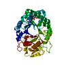
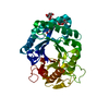
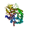
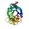
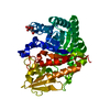
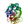
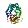
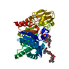
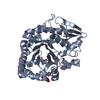
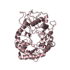
- PDB-2dt0: Crystal structure of the complex of goat signalling protein with ... -
+ Open data

- Basic information
| Entry | Database: PDB / ID: 2dt0 | ||||||||||||
|---|---|---|---|---|---|---|---|---|---|---|---|---|---|
| Title | Crystal structure of the complex of goat signalling protein with the trimer of N-acetylglucosamine at 2.45A resolution | ||||||||||||
 Components Components | Chitinase-3-like protein 1 | ||||||||||||
 Keywords Keywords | SIGNALING PROTEIN / SPG-40 / TRISACCHARIDE / COMPLEX | ||||||||||||
| Function / homology |  Function and homology information Function and homology informationresponse to interleukin-6 / activation of NF-kappaB-inducing kinase activity / chitin catabolic process / positive regulation of peptidyl-threonine phosphorylation / chitin binding / response to tumor necrosis factor / response to mechanical stimulus / lung development / response to interleukin-1 / positive regulation of interleukin-8 production ...response to interleukin-6 / activation of NF-kappaB-inducing kinase activity / chitin catabolic process / positive regulation of peptidyl-threonine phosphorylation / chitin binding / response to tumor necrosis factor / response to mechanical stimulus / lung development / response to interleukin-1 / positive regulation of interleukin-8 production / cellular response to tumor necrosis factor / positive regulation of angiogenesis / carbohydrate binding / carbohydrate metabolic process / positive regulation of ERK1 and ERK2 cascade / positive regulation of phosphatidylinositol 3-kinase/protein kinase B signal transduction / inflammatory response / apoptotic process / perinuclear region of cytoplasm / endoplasmic reticulum / : / cytoplasm Similarity search - Function | ||||||||||||
| Biological species |  | ||||||||||||
| Method |  X-RAY DIFFRACTION / MOLECULAR REPLACEMENT / Resolution: 2.45 Å X-RAY DIFFRACTION / MOLECULAR REPLACEMENT / Resolution: 2.45 Å | ||||||||||||
 Authors Authors | Kumar, J. / Ethayathulla, A.S. / Srivastava, D.B. / Singh, N. / Sharma, S. / Bhushan, A. / Srinivasan, A. / Singh, T.P. | ||||||||||||
 Citation Citation | Journal: ACTA CRYSTALLOGR.,SECT.D / Year: 2007 Title: Carbohydrate-binding properties of goat secretory glycoprotein (SPG-40) and its functional implications: structures of the native glycoprotein and its four complexes with chitin-like oligosaccharides Authors: Kumar, J. / Ethayathulla, A.S. / Srivastava, D.B. / Singh, N. / Sharma, S. / Kaur, P. / Srinivasan, A. / Singh, T.P. | ||||||||||||
| History |
|
- Structure visualization
| Structure viewer | Molecule: MolmilJmol/JSmol |
|---|
- Downloads & links
-Download
| PDBx/mmCIF format | 2dt0.cif.gz | 92.4 KB | Display | PDBx/mmCIF format |
|---|---|---|---|---|
| PDB format | pdb2dt0.ent.gz | 68.6 KB | Display | PDB format |
| PDBx/mmJSON format | 2dt0.json.gz | Tree view | PDBx/mmJSON format | |
| Others |  Other downloads Other downloads |
-Validation report
| Arichive directory | https://data.pdbj.org/pub/pdb/validation_reports/dt/2dt0ftp://data.pdbj.org/pub/pdb/validation_reports/dt/2dt0 | HTTPS FTP |
|---|
-Related structure data
| Related structure data |  2dszC 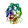 2dt1C  2dt2C 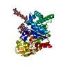 2dt3C  1ljyS C: citing same article ( S: Starting model for refinement |
|---|---|
| Similar structure data |


-Links
 PDBj
PDBj
- Assembly
| Deposited unit | 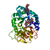
| ||||||||
|---|---|---|---|---|---|---|---|---|---|
| 1 |
| ||||||||
| Unit cell |
|
-Components
| #1: Protein | Mass: 40728.090 Da / Num. of mol.: 1 / Source method: isolated from a natural source / Source: (natural) |
|---|---|
| #2: Polysaccharide | beta-D-mannopyranose-(1-4)-2-acetamido-2-deoxy-alpha-D-glucopyranose-(1-4)-2-acetamido-2-deoxy-beta- ...beta-D-mannopyranose-(1-4)-2-acetamido-2-deoxy-alpha-D-glucopyranose-(1-4)-2-acetamido-2-deoxy-beta-D-glucopyranose Source method: isolated from a genetically manipulated source |
| #3: Polysaccharide | 2-acetamido-2-deoxy-beta-D-glucopyranose-(1-4)-2-acetamido-2-deoxy-beta-D-glucopyranose-(1-4)-2- ...2-acetamido-2-deoxy-beta-D-glucopyranose-(1-4)-2-acetamido-2-deoxy-beta-D-glucopyranose-(1-4)-2-acetamido-2-deoxy-beta-D-glucopyranose / triacetyl-beta-chitotriose  Source method: isolated from a genetically manipulated source Details: oligosaccharide / References: triacetyl-beta-chitotriose |
| #4: Water | ChemComp-HOH /  Mass: 18.015 Da / Num. of mol.: 159 / Source method: isolated from a natural source / Formula: H2O Mass: 18.015 Da / Num. of mol.: 159 / Source method: isolated from a natural source / Formula: H2O |
| Has protein modification | Y |
-Experimental details
-Experiment
| Experiment | Method: X-RAY DIFFRACTION / Number of used crystals: 1 |
|---|
- Sample preparation
| Crystal | Density Matthews: 2.76 Å3/Da / Density % sol: 55.42 % |
|---|---|
| Crystal grow | Temperature: 298 K / Method: vapor diffusion, hanging drop / pH: 7.8 Details: 25MM TRIS HCL, 50MM NACL, 19% Ethanol, pH 7.8, VAPOR DIFFUSION, HANGING DROP, temperature 298.0K |
-Data collection
| Diffraction | Mean temperature: 298 K |
|---|---|
| Diffraction source | Source: ROTATING ANODE / Type: RIGAKU RU300 / Wavelength: 1.5418 Å |
| Detector | Type: MARRESEARCH / Detector: IMAGE PLATE / Date: Jun 10, 2005 / Details: MIRROR |
| Radiation | Monochromator: GRAPHITE / Protocol: SINGLE WAVELENGTH / Monochromatic (M) / Laue (L): M / Scattering type: x-ray |
| Radiation wavelength | Wavelength: 1.5418 Å / Relative weight: 1 |
| Reflection | Resolution: 2.45→56 Å / Num. obs: 15904 / % possible obs: 98.3 % / Observed criterion σ(F): 0 / Observed criterion σ(I): 0 / Biso Wilson estimate: 44.4 Å2 / Rsym value: 0.09 / Net I/σ(I): 7 |
| Reflection shell | Resolution: 2.45→2.66 Å / Mean I/σ(I) obs: 2 / Rsym value: 0.355 / % possible all: 95 |
- Processing
| Software |
| ||||||||||||||||||||||||||||||||||||||||||||||||||||||||||||||||||||||||||||||||
|---|---|---|---|---|---|---|---|---|---|---|---|---|---|---|---|---|---|---|---|---|---|---|---|---|---|---|---|---|---|---|---|---|---|---|---|---|---|---|---|---|---|---|---|---|---|---|---|---|---|---|---|---|---|---|---|---|---|---|---|---|---|---|---|---|---|---|---|---|---|---|---|---|---|---|---|---|---|---|---|---|---|
| Refinement | Method to determine structure: MOLECULAR REPLACEMENT Starting model: 1LJY Resolution: 2.45→56 Å / Rfactor Rfree error: 0.014 / Data cutoff high absF: 1500079.47 / Data cutoff low absF: 0 / Isotropic thermal model: RESTRAINED / Cross valid method: THROUGHOUT / σ(F): 0 / σ(I): 0 / Stereochemistry target values: Engh & Huber
| ||||||||||||||||||||||||||||||||||||||||||||||||||||||||||||||||||||||||||||||||
| Solvent computation | Solvent model: FLAT MODEL / Bsol: 70.1396 Å2 / ksol: 0.343087 e/Å3 | ||||||||||||||||||||||||||||||||||||||||||||||||||||||||||||||||||||||||||||||||
| Displacement parameters | Biso mean: 44.1 Å2
| ||||||||||||||||||||||||||||||||||||||||||||||||||||||||||||||||||||||||||||||||
| Refine analyze |
| ||||||||||||||||||||||||||||||||||||||||||||||||||||||||||||||||||||||||||||||||
| Refinement step | Cycle: LAST / Resolution: 2.45→56 Å
| ||||||||||||||||||||||||||||||||||||||||||||||||||||||||||||||||||||||||||||||||
| Refine LS restraints |
| ||||||||||||||||||||||||||||||||||||||||||||||||||||||||||||||||||||||||||||||||
| LS refinement shell | Resolution: 2.45→2.66 Å / Rfactor Rfree error: 0.044 / Total num. of bins used: 6
| ||||||||||||||||||||||||||||||||||||||||||||||||||||||||||||||||||||||||||||||||
| Xplor file |
|
