 ムービー
ムービー コントローラー


+ データを開く

- 基本情報
基本情報
| 登録情報 | データベース: PDB / ID: 2dsl | ||||||
|---|---|---|---|---|---|---|---|
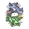
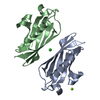
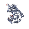
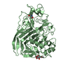
| タイトル | Mutant N33D structure of phenylacetic acid degradation protein PaaI from Thermus thermophilus HB8 | ||||||
 要素 要素 | Phenylacetic acid degradation protein PaaI | ||||||
 キーワード キーワード | HYDROLASE / THIOESTERASE / HOT DOG FOLD / PHENYLACETIC ACID DEGRADATION / Structural Genomics / NPPSFA / National Project on Protein Structural and Functional Analyses / RIKEN Structural Genomics/Proteomics Initiative / RSGI | ||||||
| 機能・相同性 |  機能・相同性情報 機能・相同性情報 | ||||||
| 生物種 |   Thermus thermophilus (バクテリア) Thermus thermophilus (バクテリア) | ||||||
| 手法 |  X線回折 / 分子置換 / 解像度: 1.7 Å X線回折 / 分子置換 / 解像度: 1.7 Å | ||||||
 データ登録者 データ登録者 | Shimizu, K. / RIKEN Structural Genomics/Proteomics Initiative (RSGI) | ||||||
 引用 引用 | ジャーナル: To be Published タイトル: Mutant N33D structure of phenylacetic acid degradation protein PaaI from Thermus thermophilus HB8 著者: Shimizu, K. / Sugahara, M. / Kunishima, N. | ||||||
| 履歴 |
|
- 構造の表示
構造の表示
| 構造ビューア | 分子: MolmilJmol/JSmol |
|---|
- ダウンロードとリンク
-ダウンロード
| PDBx/mmCIF形式 | 2dsl.cif.gz | 61.3 KB | 表示 | PDBx/mmCIF形式 |
|---|---|---|---|---|
| PDB形式 | pdb2dsl.ent.gz | 44 KB | 表示 | PDB形式 |
| PDBx/mmJSON形式 | 2dsl.json.gz | ツリー表示 | PDBx/mmJSON形式 | |
| その他 |  その他のダウンロード その他のダウンロード |
-検証レポート
| アーカイブディレクトリ | https://data.pdbj.org/pub/pdb/validation_reports/ds/2dslftp://data.pdbj.org/pub/pdb/validation_reports/ds/2dsl | HTTPS FTP |
|---|
-関連構造データ
| 関連構造データ |  1j1yS S: 精密化の開始モデル |
|---|---|
| 類似構造データ | |
| その他のデータベース |


-リンク
 PDBj
PDBj
- 集合体
集合体
| 登録構造単位 | 
| |||||||||||||||||||||
|---|---|---|---|---|---|---|---|---|---|---|---|---|---|---|---|---|---|---|---|---|---|---|
| 1 | 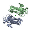
| |||||||||||||||||||||
| 単位格子 |
| |||||||||||||||||||||
| Components on special symmetry positions |
| |||||||||||||||||||||
| 詳細 | The biological assembly is a tetramer generated from the dimer A and B in the asymmetric unit. |
-要素
| #1: タンパク質 | 分子量: 14281.208 Da / 分子数: 2 / 変異: N33D / 由来タイプ: 組換発現 由来: (組換発現) Thermus thermophilus (バクテリア)株: HB8 / プラスミド: pET11a / 生物種 (発現宿主): Escherichia coli / 発現宿主: #2: 化合物 | ChemComp-CL / |   分子量: 35.453 Da / 分子数: 1 / 由来タイプ: 合成 / 式: Cl 分子量: 35.453 Da / 分子数: 1 / 由来タイプ: 合成 / 式: Cl#3: 化合物 | ChemComp-MG / |   分子量: 24.305 Da / 分子数: 1 / 由来タイプ: 合成 / 式: Mg 分子量: 24.305 Da / 分子数: 1 / 由来タイプ: 合成 / 式: Mg#4: 水 | ChemComp-HOH / |  分子量: 18.015 Da / 分子数: 233 / 由来タイプ: 天然 / 式: H2O 分子量: 18.015 Da / 分子数: 233 / 由来タイプ: 天然 / 式: H2O |
|---|
-実験情報
-実験
| 実験 | 手法: X線回折 / 使用した結晶の数: 1 |
|---|
- 試料調製
試料調製
| 結晶 | マシュー密度: 2.03 Å3/Da / 溶媒含有率: 39.48 % |
|---|---|
| 結晶化 | 温度: 295 K / 手法: microbach / pH: 7.5 詳細: 20% PEG 400, 0.2M Magnesium Chloride, 0.1M HEPES, pH 7.5, Microbach, temperature 295K |
-データ収集
| 回折 | 平均測定温度: 100 K |
|---|---|
| 放射光源 | 由来: 回転陽極 / タイプ: RIGAKU / 波長: 1.5418 Å |
| 検出器 | タイプ: RIGAKU RAXIS V / 検出器: IMAGE PLATE / 日付: 2002年7月23日 / 詳細: Mirror |
| 放射 | モノクロメーター: Mirror / プロトコル: SINGLE WAVELENGTH / 単色(M)・ラウエ(L): M / 散乱光タイプ: x-ray |
| 放射波長 | 波長: 1.5418 Å / 相対比: 1 |
| 反射 | 解像度: 1.7→30 Å / Num. all: 26557 / Num. obs: 26557 / % possible obs: 98.7 % / Observed criterion σ(F): 0 / Observed criterion σ(I): 0 / 冗長度: 6.39 % / Biso Wilson estimate: 25.1 Å2 / Rmerge(I) obs: 0.059 / Rsym value: 0.056 / Net I/σ(I): 12.2 |
| 反射 シェル | 解像度: 1.7→1.76 Å / 冗長度: 6.55 % / Rmerge(I) obs: 0.412 / Mean I/σ(I) obs: 3.3 / Num. unique all: 26557 / Rsym value: 0.384 / % possible all: 99.3 |
- 解析
解析
| ソフトウェア |
| ||||||||||||||||||||||||||||||||||||
|---|---|---|---|---|---|---|---|---|---|---|---|---|---|---|---|---|---|---|---|---|---|---|---|---|---|---|---|---|---|---|---|---|---|---|---|---|---|
| 精密化 | 構造決定の手法: 分子置換 開始モデル: PDB ENTRY 1J1Y 解像度: 1.7→22.56 Å / Rfactor Rfree error: 0.006 / Data cutoff high absF: 1626023.85 / Data cutoff low absF: 0 / Isotropic thermal model: RESTRAINED / 交差検証法: THROUGHOUT / σ(F): 0 / σ(I): 0 / 立体化学のターゲット値: Engh & Huber
| ||||||||||||||||||||||||||||||||||||
| 溶媒の処理 | 溶媒モデル: FLAT MODEL / Bsol: 61.115 Å2 / ksol: 0.393909 e/Å3 | ||||||||||||||||||||||||||||||||||||
| 原子変位パラメータ | Biso mean: 24.6 Å2
| ||||||||||||||||||||||||||||||||||||
| Refine analyze |
| ||||||||||||||||||||||||||||||||||||
| 精密化ステップ | サイクル: LAST / 解像度: 1.7→22.56 Å
| ||||||||||||||||||||||||||||||||||||
| 拘束条件 |
| ||||||||||||||||||||||||||||||||||||
| LS精密化 シェル | 解像度: 1.7→1.81 Å / Rfactor Rfree error: 0.021 / Total num. of bins used: 6
| ||||||||||||||||||||||||||||||||||||
| Xplor file |
|
