 Movie
Movie Controller

[English] 日本語
 Yorodumi
- PDB-2djg: Re-determination of the native structure of human dipeptidyl pept... -
+ Open data

- Basic information
| Entry | Database: PDB / ID: 2djg | |||||||||
|---|---|---|---|---|---|---|---|---|---|---|
| Title | Re-determination of the native structure of human dipeptidyl peptidase I (cathepsin C) | |||||||||
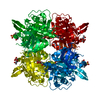
 Components Components | (Dipeptidyl-peptidase ...) x 3 | |||||||||
 Keywords Keywords | HYDROLASE / re-refinement / cysteine protease / cathepsin C / dipeptidyl peptidase I | |||||||||
| Function / homology |  Function and homology information Function and homology informationdipeptidyl-peptidase I / peptidase activator activity involved in apoptotic process / : / Cargo concentration in the ER / COPII-coated ER to Golgi transport vesicle / dipeptidyl-peptidase activity / COPII-mediated vesicle transport / chloride ion binding / phosphatase binding / endoplasmic reticulum-Golgi intermediate compartment membrane ...dipeptidyl-peptidase I / peptidase activator activity involved in apoptotic process / : / Cargo concentration in the ER / COPII-coated ER to Golgi transport vesicle / dipeptidyl-peptidase activity / COPII-mediated vesicle transport / chloride ion binding / phosphatase binding / endoplasmic reticulum-Golgi intermediate compartment membrane / cysteine-type peptidase activity / MHC class II antigen presentation / : / T cell mediated cytotoxicity / azurophil granule lumen / protein-folding chaperone binding / extracellular matrix / lysosome / immune response / endoplasmic reticulum lumen / serine-type endopeptidase activity / cysteine-type endopeptidase activity / Neutrophil degranulation / proteolysis / : / extracellular exosome / extracellular region / membrane / identical protein binding Similarity search - Function | |||||||||
| Biological species |  Homo sapiens (human) Homo sapiens (human) | |||||||||
| Method |  X-RAY DIFFRACTION / MOLECULAR REPLACEMENT / Resolution: 2.05 Å X-RAY DIFFRACTION / MOLECULAR REPLACEMENT / Resolution: 2.05 Å | |||||||||
 Authors Authors | Molgaard, A. / Arnau, J. / Lauritzen, C. / Larsen, S. / Petersen, G. / Pedersen, J. | |||||||||
 Citation Citation | Journal: Biochem.J. / Year: 2007 Title: The crystal structure of human dipeptidyl peptidase I (cathepsin C) in complex with the inhibitor Gly-Phe-CHN2 Authors: Molgaard, A. / Arnau, J. / Lauritzen, C. / Larsen, S. / Petersen, G. / Pedersen, J. | |||||||||
| History |
|
- Structure visualization
| Structure viewer | Molecule: MolmilJmol/JSmol |
|---|
- Downloads & links
-Download
| PDBx/mmCIF format | 2djg.cif.gz | 90.3 KB | Display | PDBx/mmCIF format |
|---|---|---|---|---|
| PDB format | pdb2djg.ent.gz | 67.2 KB | Display | PDB format |
| PDBx/mmJSON format | 2djg.json.gz | Tree view | PDBx/mmJSON format | |
| Others |  Other downloads Other downloads |
-Validation report
| Arichive directory | https://data.pdbj.org/pub/pdb/validation_reports/dj/2djgftp://data.pdbj.org/pub/pdb/validation_reports/dj/2djg | HTTPS FTP |
|---|
-Related structure data
| Related structure data | 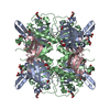 2djfC  1k3bS S: Starting model for refinement C: citing same article ( |
|---|---|
| Similar structure data |


-Links
 PDBj
PDBj


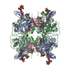
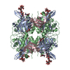
- Assembly
| Deposited unit | 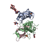
| ||||||||
|---|---|---|---|---|---|---|---|---|---|
| 1 | 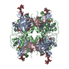
| ||||||||
| Unit cell |
|
-Components
-Dipeptidyl-peptidase ... , 3 types, 3 molecules ABC
| #1: Protein | Mass: 13500.163 Da / Num. of mol.: 1 / Fragment: Dipeptidyl-peptidase 1 exclusion domain chain Source method: isolated from a genetically manipulated source Source: (gene. exp.) Homo sapiens (human) / Gene: CTSC / Cell (production host): BTI-TN-5B1-4 / Production host:  Trichoplusia ni (cabbage looper) / References: UniProt: P53634, dipeptidyl-peptidase I Trichoplusia ni (cabbage looper) / References: UniProt: P53634, dipeptidyl-peptidase I |
|---|---|
| #2: Protein | Mass: 18491.871 Da / Num. of mol.: 1 / Fragment: Dipeptidyl-peptidase 1 heavy chain Source method: isolated from a genetically manipulated source Source: (gene. exp.) Homo sapiens (human) / Gene: CTSC / Cell (production host): BTI-TN-5B1-4 / Production host: Trichoplusia ni (cabbage looper) / References: UniProt: P53634, dipeptidyl-peptidase I |
| #3: Protein | Mass: 7583.444 Da / Num. of mol.: 1 / Fragment: Dipeptidyl-peptidase 1 light chain Source method: isolated from a genetically manipulated source Source: (gene. exp.) Homo sapiens (human) / Gene: CTSC / Cell (production host): BTI-TN-5B1-4 / Production host: Trichoplusia ni (cabbage looper) / References: UniProt: P53634, dipeptidyl-peptidase I |
-Sugars , 2 types, 3 molecules 
| #4: Polysaccharide | beta-D-mannopyranose-(1-2)-beta-D-mannopyranose-(1-3)-beta-D-mannopyranose-(1-4)-2-acetamido-2- ...beta-D-mannopyranose-(1-2)-beta-D-mannopyranose-(1-3)-beta-D-mannopyranose-(1-4)-2-acetamido-2-deoxy-beta-D-glucopyranose-(1-4)-2-acetamido-2-deoxy-beta-D-glucopyranose Source method: isolated from a genetically manipulated source |
|---|---|
| #5: Sugar |  Type: D-saccharide, beta linking / Mass: 221.208 Da / Num. of mol.: 2 Type: D-saccharide, beta linking / Mass: 221.208 Da / Num. of mol.: 2Source method: isolated from a genetically manipulated source Formula: C8H15NO6 |
-Non-polymers , 3 types, 214 molecules 


| #6: Chemical |  Mass: 96.063 Da / Num. of mol.: 2 / Source method: obtained synthetically / Formula: SO4 Mass: 96.063 Da / Num. of mol.: 2 / Source method: obtained synthetically / Formula: SO4#7: Chemical | ChemComp-CL / |  Mass: 35.453 Da / Num. of mol.: 1 / Source method: obtained synthetically / Formula: Cl Mass: 35.453 Da / Num. of mol.: 1 / Source method: obtained synthetically / Formula: Cl#8: Water | ChemComp-HOH / | Mass: 18.015 Da / Num. of mol.: 211 / Source method: isolated from a natural source / Formula: H2O |
|---|
-Details
| Has protein modification | Y |
|---|
-Experimental details
-Experiment
| Experiment | Method: X-RAY DIFFRACTION / Number of used crystals: 1 |
|---|
- Sample preparation
| Crystal | Density Matthews: 2.8 Å3/Da / Density % sol: 56.08 % |
|---|---|
| Crystal grow | Temperature: 298 K / Method: vapor diffusion, hanging drop / pH: 5.4 Details: 1.8M ammonium sulfate, 0.1M Na citrate, 0.2M Na/K tartrate, pH 5.4, VAPOR DIFFUSION, HANGING DROP, temperature 298K |
-Data collection
| Diffraction | Mean temperature: 120 K |
|---|---|
| Diffraction source | Source: ROTATING ANODE / Type: RIGAKU RU300 / Wavelength: 1.5418 Å |
| Detector | Type: MAR scanner 345 mm plate / Detector: IMAGE PLATE / Date: Aug 23, 2004 |
| Radiation | Protocol: SINGLE WAVELENGTH / Monochromatic (M) / Laue (L): M / Scattering type: x-ray |
| Radiation wavelength | Wavelength: 1.5418 Å / Relative weight: 1 |
| Reflection | Resolution: 2.05→30 Å / Num. all: 120690 / Num. obs: 120690 |
| Reflection shell | Resolution: 2.05→2.12 Å / % possible all: 96.1 |
- Processing
| Software |
| ||||||||||||||||||||||||||||||||||||||||||||||||||||||||||||||||||||||||||||||||||||||||||||||||||||||||||||||||||||||||||||||||||||||||||||||||||||||||||||||||||||||||||
|---|---|---|---|---|---|---|---|---|---|---|---|---|---|---|---|---|---|---|---|---|---|---|---|---|---|---|---|---|---|---|---|---|---|---|---|---|---|---|---|---|---|---|---|---|---|---|---|---|---|---|---|---|---|---|---|---|---|---|---|---|---|---|---|---|---|---|---|---|---|---|---|---|---|---|---|---|---|---|---|---|---|---|---|---|---|---|---|---|---|---|---|---|---|---|---|---|---|---|---|---|---|---|---|---|---|---|---|---|---|---|---|---|---|---|---|---|---|---|---|---|---|---|---|---|---|---|---|---|---|---|---|---|---|---|---|---|---|---|---|---|---|---|---|---|---|---|---|---|---|---|---|---|---|---|---|---|---|---|---|---|---|---|---|---|---|---|---|---|---|---|---|
| Refinement | Method to determine structure: MOLECULAR REPLACEMENT Starting model: 1k3b Resolution: 2.05→24.04 Å / Cor.coef. Fo:Fc: 0.954 / Cor.coef. Fo:Fc free: 0.931 / SU B: 3.62 / SU ML: 0.099 / Cross valid method: THROUGHOUT / ESU R: 0.173 / ESU R Free: 0.16 / Stereochemistry target values: MAXIMUM LIKELIHOOD / Details: HYDROGENS HAVE BEEN ADDED IN THE RIDING POSITIONS
| ||||||||||||||||||||||||||||||||||||||||||||||||||||||||||||||||||||||||||||||||||||||||||||||||||||||||||||||||||||||||||||||||||||||||||||||||||||||||||||||||||||||||||
| Solvent computation | Ion probe radii: 0.8 Å / Shrinkage radii: 0.8 Å / VDW probe radii: 1.4 Å / Solvent model: MASK | ||||||||||||||||||||||||||||||||||||||||||||||||||||||||||||||||||||||||||||||||||||||||||||||||||||||||||||||||||||||||||||||||||||||||||||||||||||||||||||||||||||||||||
| Displacement parameters | Biso mean: 23.055 Å2
| ||||||||||||||||||||||||||||||||||||||||||||||||||||||||||||||||||||||||||||||||||||||||||||||||||||||||||||||||||||||||||||||||||||||||||||||||||||||||||||||||||||||||||
| Refinement step | Cycle: LAST / Resolution: 2.05→24.04 Å
| ||||||||||||||||||||||||||||||||||||||||||||||||||||||||||||||||||||||||||||||||||||||||||||||||||||||||||||||||||||||||||||||||||||||||||||||||||||||||||||||||||||||||||
| Refine LS restraints |
| ||||||||||||||||||||||||||||||||||||||||||||||||||||||||||||||||||||||||||||||||||||||||||||||||||||||||||||||||||||||||||||||||||||||||||||||||||||||||||||||||||||||||||
| LS refinement shell | Resolution: 2.05→2.103 Å / Total num. of bins used: 20 /
|
