 Movie
Movie Controller

[English] 日本語
 Yorodumi
- PDB-2df3: The structure of Siglec-7 in complex with alpha(2,3)/alpha(2,6) d... -
+ Open data

- Basic information
| Entry | Database: PDB / ID: 2df3 | |||||||||||||||
|---|---|---|---|---|---|---|---|---|---|---|---|---|---|---|---|---|
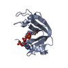
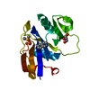
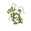
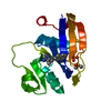
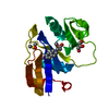
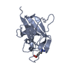
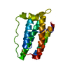
| Title | The structure of Siglec-7 in complex with alpha(2,3)/alpha(2,6) disialyl lactotetraosyl 2-(trimethylsilyl)ethyl | |||||||||||||||
 Components Components | Sialic acid-binding Ig-like lectin 7 | |||||||||||||||
 Keywords Keywords | CELL ADHESION / Siglec / sialic acid / ganglioside | |||||||||||||||
| Function / homology |  Function and homology information Function and homology informationsialic acid binding / Immunoregulatory interactions between a Lymphoid and a non-Lymphoid cell / carbohydrate binding / signaling receptor activity / cell adhesion / plasma membrane Similarity search - Function | |||||||||||||||
| Biological species |  Homo sapiens (human) Homo sapiens (human) | |||||||||||||||
| Method |  X-RAY DIFFRACTION / SYNCHROTRON / MOLECULAR REPLACEMENT / Resolution: 1.9 Å X-RAY DIFFRACTION / SYNCHROTRON / MOLECULAR REPLACEMENT / Resolution: 1.9 Å | |||||||||||||||
 Authors Authors | Attrill, H. | |||||||||||||||
 Citation Citation | Journal: Biochem.J. / Year: 2006 Title: The structure of siglec-7 in complex with sialosides: leads for rational structure-based inhibitor design Authors: Attrill, H. / Takazawa, H. / Witt, S. / Kelm, S. / Isecke, R. / Brossmer, R. / Ando, T. / Ishida, H. / Kiso, M. / Crocker, P.R. / van Aalten, D.M.F. | |||||||||||||||
| History |
|
- Structure visualization
| Structure viewer | Molecule: MolmilJmol/JSmol |
|---|
- Downloads & links
-Download
| PDBx/mmCIF format | 2df3.cif.gz | 43.9 KB | Display | PDBx/mmCIF format |
|---|---|---|---|---|
| PDB format | pdb2df3.ent.gz | 28.8 KB | Display | PDB format |
| PDBx/mmJSON format | 2df3.json.gz | Tree view | PDBx/mmJSON format | |
| Others |  Other downloads Other downloads |
-Validation report
| Arichive directory | https://data.pdbj.org/pub/pdb/validation_reports/df/2df3ftp://data.pdbj.org/pub/pdb/validation_reports/df/2df3 | HTTPS FTP |
|---|
-Related structure data
| Related structure data | 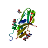 2g5rC  1o7sS S: Starting model for refinement C: citing same article ( |
|---|---|
| Similar structure data |


-Links
 PDBj
PDBj


- Assembly
| Deposited unit | 
| ||||||||
|---|---|---|---|---|---|---|---|---|---|
| 1 |
| ||||||||
| Unit cell |
|
-Components
| #1: Protein | Mass: 14655.292 Da / Num. of mol.: 1 / Fragment: Ig-like V-type Source method: isolated from a genetically manipulated source Source: (gene. exp.) Homo sapiens (human) / Gene: SIGLEC7, AIRM1 / Plasmid: pDEF / Cell line (production host): CHO Lec1 / Production host:   Cricetulus griseus (Chinese hamster) / References: UniProt: Q9Y286 Cricetulus griseus (Chinese hamster) / References: UniProt: Q9Y286 |
|---|---|
| #2: Polysaccharide | N-acetyl-alpha-neuraminic acid-(2-3)-beta-D-galactopyranose-(1-3)-[N-acetyl-alpha-neuraminic acid- ...N-acetyl-alpha-neuraminic acid-(2-3)-beta-D-galactopyranose-(1-3)-[N-acetyl-alpha-neuraminic acid-(2-6)]2-acetamido-2-deoxy-beta-D-glucopyranose Source method: isolated from a genetically manipulated source |
| #3: Sugar | ChemComp-NAG /   Type: D-saccharide, beta linking / Mass: 221.208 Da / Num. of mol.: 1 Type: D-saccharide, beta linking / Mass: 221.208 Da / Num. of mol.: 1Source method: isolated from a genetically manipulated source Formula: C8H15NO6 |
| #4: Chemical | ChemComp-CYS /   Type: L-peptide linking / Mass: 121.158 Da / Num. of mol.: 1 / Source method: obtained synthetically / Formula: C3H7NO2S Type: L-peptide linking / Mass: 121.158 Da / Num. of mol.: 1 / Source method: obtained synthetically / Formula: C3H7NO2S |
| #5: Water | ChemComp-HOH /  Mass: 18.015 Da / Num. of mol.: 94 / Source method: isolated from a natural source / Formula: H2O Mass: 18.015 Da / Num. of mol.: 94 / Source method: isolated from a natural source / Formula: H2O |
| Has protein modification | Y |
-Experimental details
-Experiment
| Experiment | Method: X-RAY DIFFRACTION / Number of used crystals: 1 |
|---|
- Sample preparation
| Crystal | Density Matthews: 2.23 Å3/Da / Density % sol: 44.84 % |
|---|---|
| Crystal grow | Temperature: 293 K / Method: vapor diffusion, sitting drop / pH: 7.5 Details: 8.5% iso-propanol, 0.095M HEPES, 17% PEG 4000, 15% glycerol, pH 7.5, VAPOR DIFFUSION, SITTING DROP, temperature 293K |
-Data collection
| Diffraction | Mean temperature: 100 K |
|---|---|
| Diffraction source | Source: SYNCHROTRON / Site: ESRF  / Beamline: ID14-4 / Wavelength: 0.933 Å / Beamline: ID14-4 / Wavelength: 0.933 Å |
| Detector | Type: ADSC QUANTUM 4 / Detector: CCD / Date: Feb 2, 2003 |
| Radiation | Protocol: SINGLE WAVELENGTH / Monochromatic (M) / Laue (L): M / Scattering type: x-ray |
| Radiation wavelength | Wavelength: 0.933 Å / Relative weight: 1 |
| Reflection | Resolution: 1.9→25 Å / Num. obs: 11014 / % possible obs: 24 % / Observed criterion σ(F): 2 / Observed criterion σ(I): 2 / Redundancy: 4.3 % / Net I/σ(I): 15.5 |
| Reflection shell | Resolution: 1.9→1.97 Å / Redundancy: 3.8 % / Rmerge(I) obs: 0.38 / Mean I/σ(I) obs: 3.1 / Num. unique all: 1011 / % possible all: 20 |
- Processing
| Software |
| ||||||||||||||||||||||||||||||||||||||||||||||||||||||||||||||||||||||
|---|---|---|---|---|---|---|---|---|---|---|---|---|---|---|---|---|---|---|---|---|---|---|---|---|---|---|---|---|---|---|---|---|---|---|---|---|---|---|---|---|---|---|---|---|---|---|---|---|---|---|---|---|---|---|---|---|---|---|---|---|---|---|---|---|---|---|---|---|---|---|---|
| Refinement | Method to determine structure: MOLECULAR REPLACEMENT Starting model: PDB ENTRY 1O7S Resolution: 1.9→25 Å / Cor.coef. Fo:Fc: 0.951 / Cor.coef. Fo:Fc free: 0.917 / SU B: 3.401 / SU ML: 0.098 / Cross valid method: THROUGHOUT / σ(F): 0 / ESU R: 0.164 / ESU R Free: 0.15 / Stereochemistry target values: MAXIMUM LIKELIHOOD
| ||||||||||||||||||||||||||||||||||||||||||||||||||||||||||||||||||||||
| Solvent computation | Ion probe radii: 0.8 Å / Shrinkage radii: 0.8 Å / VDW probe radii: 1.4 Å / Solvent model: BABINET MODEL WITH MASK | ||||||||||||||||||||||||||||||||||||||||||||||||||||||||||||||||||||||
| Displacement parameters | Biso mean: 20.857 Å2
| ||||||||||||||||||||||||||||||||||||||||||||||||||||||||||||||||||||||
| Refinement step | Cycle: LAST / Resolution: 1.9→25 Å
| ||||||||||||||||||||||||||||||||||||||||||||||||||||||||||||||||||||||
| Refine LS restraints |
| ||||||||||||||||||||||||||||||||||||||||||||||||||||||||||||||||||||||
| LS refinement shell | Resolution: 1.9→1.949 Å / Total num. of bins used: 20
|
