 Movie
Movie Controller

[English] 日本語
 Yorodumi
- PDB-2dci: NMR structure of influenza HA fusion peptide mutant W14A in DPC in pH5 -
+ Open data

- Basic information
| Entry | Database: PDB / ID: 2dci | ||||||
|---|---|---|---|---|---|---|---|
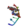
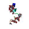
| Title | NMR structure of influenza HA fusion peptide mutant W14A in DPC in pH5 | ||||||
 Components Components | Hemagglutinin | ||||||
 Keywords Keywords | VIRAL PROTEIN / HA / fusion peptide | ||||||
| Function / homology |  Function and homology information Function and homology informationclathrin-dependent endocytosis of virus by host cell / host cell surface receptor binding / fusion of virus membrane with host plasma membrane / fusion of virus membrane with host endosome membrane / viral envelope / virion attachment to host cell / host cell plasma membrane / virion membrane Similarity search - Function | ||||||
| Method | SOLUTION NMR / distance geometry | ||||||
 Authors Authors | Tamm, L.K. / Lai, A.L. | ||||||
 Citation Citation | Journal: J.Biol.Chem. / Year: 2006 Title: Fusion peptide of influenza hemagglutinin requires a fixed angle boomerang structure for activity Authors: Lai, A.L. / Park, H. / White, J.M. / Tamm, L.K. | ||||||
| History |
|
- Structure visualization
| Structure viewer | Molecule: MolmilJmol/JSmol |
|---|
- Downloads & links
-Download
| PDBx/mmCIF format | 2dci.cif.gz | 152.2 KB | Display | PDBx/mmCIF format |
|---|---|---|---|---|
| PDB format | pdb2dci.ent.gz | 106.6 KB | Display | PDB format |
| PDBx/mmJSON format | 2dci.json.gz | Tree view | PDBx/mmJSON format | |
| Others |  Other downloads Other downloads |
-Validation report
| Arichive directory | https://data.pdbj.org/pub/pdb/validation_reports/dc/2dciftp://data.pdbj.org/pub/pdb/validation_reports/dc/2dci | HTTPS FTP |
|---|
-Related structure data
| Related structure data | |
|---|---|
| Similar structure data |


-Links
 PDBj
PDBj


- Assembly
| Deposited unit | 
| |||||||||
|---|---|---|---|---|---|---|---|---|---|---|
| 1 |
| |||||||||
| NMR ensembles |
|
-Components
| #1: Protein/peptide | Mass: 1939.152 Da / Num. of mol.: 1 / Source method: obtained synthetically Details: Derived from the influenza (X11) hemagglutinin HA2, residue 1-20 except the 14th residue W was replaced with A. References: UniProt: P11134 |
|---|
-Experimental details
-Experiment
| Experiment | Method: SOLUTION NMR | ||||||||||||
|---|---|---|---|---|---|---|---|---|---|---|---|---|---|
| NMR experiment |
| ||||||||||||
| NMR details | Text: This structure was determined using standard 2D homonuclear techniques. |
- Sample preparation
| Details | Contents: 2mM peptide, 400mM d38-DPC, 5mM dtt, 20 mMd4-acetic acid, pH 5; 95% H2O, 5% D2O Solvent system: 95% H2O/5% D2O |
|---|---|
| Sample conditions | Ionic strength: 0 / pH: 5 / Pressure: 1 atm / Temperature: 303 K |
-NMR measurement
| NMR spectrometer | Type: Varian INOVA / Manufacturer: Varian / Model: INOVA / Field strength: 600 MHz |
|---|
- Processing
| NMR software | Name: OPAL Developer: Luginbuhl, P., Guntert, P., Billeter, M. & Wuthrich K. Classification: refinement |
|---|---|
| Refinement | Method: distance geometry / Software ordinal: 1 |
| NMR representative | Selection criteria: closest to the average |
| NMR ensemble | Conformer selection criteria: structures with the lowest energy Conformers calculated total number: 200 / Conformers submitted total number: 33 |
