 Movie
Movie Controller

[English] 日本語
 Yorodumi
- PDB-2dbw: Crystal Structure of Gamma-glutamyltranspeptidase from Escherichi... -
+ Open data

- Basic information
| Entry | Database: PDB / ID: 2dbw | ||||||
|---|---|---|---|---|---|---|---|
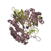
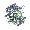
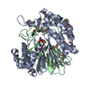
| Title | Crystal Structure of Gamma-glutamyltranspeptidase from Escherichia coli Acyl-Enzyme Intermediate | ||||||
 Components Components | (Gamma-glutamyltranspeptidase) x 2 | ||||||
 Keywords Keywords | TRANSFERASE / gamma-glutamyltransferase / ggt / gamma-gtp / glutathione / acyl-enzyme intermediate | ||||||
| Function / homology |  Function and homology information Function and homology informationamino acid salvage / gamma-glutamyl-peptidase activity / gamma-glutamyltransferase / glutathione gamma-glutamate hydrolase / glutathione gamma-glutamate hydrolase / : / glutathione catabolic process / glutathione biosynthetic process / self proteolysis / outer membrane-bounded periplasmic space / periplasmic space Similarity search - Function | ||||||
| Biological species |  | ||||||
| Method |  X-RAY DIFFRACTION / SYNCHROTRON / FOURIER SYNTHESIS / Resolution: 1.8 Å X-RAY DIFFRACTION / SYNCHROTRON / FOURIER SYNTHESIS / Resolution: 1.8 Å | ||||||
 Authors Authors | Okada, T. / Wada, K. / Fukuyama, K. | ||||||
 Citation Citation | Journal: Proc.Natl.Acad.Sci.USA / Year: 2006 Title: Crystal structures of gamma-glutamyltranspeptidase from Escherichia coli, a key enzyme in glutathione metabolism, and its reaction intermediate Authors: Okada, T. / Suzuki, H. / Wada, K. / Kumagai, H. / Fukuyama, K. | ||||||
| History |
|
- Structure visualization
| Structure viewer | Molecule: MolmilJmol/JSmol |
|---|
- Downloads & links
-Download
| PDBx/mmCIF format | 2dbw.cif.gz | 237.4 KB | Display | PDBx/mmCIF format |
|---|---|---|---|---|
| PDB format | pdb2dbw.ent.gz | 188.1 KB | Display | PDB format |
| PDBx/mmJSON format | 2dbw.json.gz | Tree view | PDBx/mmJSON format | |
| Others |  Other downloads Other downloads |
-Validation report
| Arichive directory | https://data.pdbj.org/pub/pdb/validation_reports/db/2dbwftp://data.pdbj.org/pub/pdb/validation_reports/db/2dbw | HTTPS FTP |
|---|
-Related structure data
| Related structure data |  2dbuSC  2dbxC  2dg5C S: Starting model for refinement C: citing same article ( |
|---|---|
| Similar structure data |

-Links
 PDBj
PDBj


- Assembly
| Deposited unit | 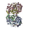
| ||||||||
|---|---|---|---|---|---|---|---|---|---|
| 1 | 
| ||||||||
| 2 | 
| ||||||||
| Unit cell |
|
-Components
| #1: Protein | Mass: 39827.070 Da / Num. of mol.: 2 / Fragment: LARGE SUBUNIT Source method: isolated from a genetically manipulated source Source: (gene. exp.) #2: Protein | Mass: 20262.951 Da / Num. of mol.: 2 / Fragment: SMALL SUBUNIT Source method: isolated from a genetically manipulated source Source: (gene. exp.) #3: Chemical | ChemComp-GOL /   Mass: 92.094 Da / Num. of mol.: 4 / Source method: obtained synthetically / Formula: C3H8O3 Mass: 92.094 Da / Num. of mol.: 4 / Source method: obtained synthetically / Formula: C3H8O3#4: Chemical |   Type: L-gamma-peptide, C-delta linking / Mass: 147.129 Da / Num. of mol.: 2 / Source method: obtained synthetically / Formula: C5H9NO4 Type: L-gamma-peptide, C-delta linking / Mass: 147.129 Da / Num. of mol.: 2 / Source method: obtained synthetically / Formula: C5H9NO4#5: Water | ChemComp-HOH / |  Mass: 18.015 Da / Num. of mol.: 896 / Source method: isolated from a natural source / Formula: H2O Mass: 18.015 Da / Num. of mol.: 896 / Source method: isolated from a natural source / Formula: H2O |
|---|
-Experimental details
-Experiment
| Experiment | Method: X-RAY DIFFRACTION / Number of used crystals: 1 |
|---|
- Sample preparation
| Crystal | Density Matthews: 2.68 Å3/Da / Density % sol: 54.03 % |
|---|---|
| Crystal grow | Temperature: 277 K / Method: vapor diffusion, hanging drop / pH: 5 Details: 12.5% PEG 4000, 0.2M magnesium sulfate, 5% glycerol, pH 5.0, VAPOR DIFFUSION, HANGING DROP, temperature 277K |
-Data collection
| Diffraction | Mean temperature: 100 K |
|---|---|
| Diffraction source | Source: SYNCHROTRON / Site: SPring-8  / Beamline: BL41XU / Wavelength: 1 Å / Beamline: BL41XU / Wavelength: 1 Å |
| Detector | Type: ADSC QUANTUM 4 / Detector: CCD / Date: Nov 8, 2005 |
| Radiation | Monochromator: SI(111) DOUBLE MONOCHROMATER / Protocol: SINGLE WAVELENGTH / Monochromatic (M) / Laue (L): M / Scattering type: x-ray |
| Radiation wavelength | Wavelength: 1 Å / Relative weight: 1 |
| Reflection | Resolution: 1.8→50 Å / Num. all: 120057 / Num. obs: 120057 / % possible obs: 100 % / Observed criterion σ(F): 1 / Observed criterion σ(I): 1 / Redundancy: 8 % / Biso Wilson estimate: 16.7 Å2 / Rmerge(I) obs: 0.063 |
| Reflection shell | Resolution: 1.8→1.86 Å / Redundancy: 8 % / Rmerge(I) obs: 0.348 / % possible all: 100 |
- Processing
| Software |
| ||||||||||||||||||||||||||||||||||||||||||||||||||||||||||||||||||||||||||||||||
|---|---|---|---|---|---|---|---|---|---|---|---|---|---|---|---|---|---|---|---|---|---|---|---|---|---|---|---|---|---|---|---|---|---|---|---|---|---|---|---|---|---|---|---|---|---|---|---|---|---|---|---|---|---|---|---|---|---|---|---|---|---|---|---|---|---|---|---|---|---|---|---|---|---|---|---|---|---|---|---|---|---|
| Refinement | Method to determine structure: FOURIER SYNTHESIS Starting model: PDB ENTRY 2DBU Resolution: 1.8→46.42 Å / Rfactor Rfree error: 0.003 / Data cutoff high absF: 2686055.04 / Data cutoff low absF: 0 / Isotropic thermal model: RESTRAINED / Cross valid method: THROUGHOUT / σ(F): 0 / Stereochemistry target values: Engh & Huber
| ||||||||||||||||||||||||||||||||||||||||||||||||||||||||||||||||||||||||||||||||
| Solvent computation | Solvent model: FLAT MODEL / Bsol: 46.016 Å2 / ksol: 0.36887 e/Å3 | ||||||||||||||||||||||||||||||||||||||||||||||||||||||||||||||||||||||||||||||||
| Displacement parameters | Biso mean: 21.9 Å2
| ||||||||||||||||||||||||||||||||||||||||||||||||||||||||||||||||||||||||||||||||
| Refine analyze |
| ||||||||||||||||||||||||||||||||||||||||||||||||||||||||||||||||||||||||||||||||
| Refinement step | Cycle: LAST / Resolution: 1.8→46.42 Å
| ||||||||||||||||||||||||||||||||||||||||||||||||||||||||||||||||||||||||||||||||
| Refine LS restraints |
| ||||||||||||||||||||||||||||||||||||||||||||||||||||||||||||||||||||||||||||||||
| LS refinement shell | Resolution: 1.8→1.91 Å / Rfactor Rfree error: 0.008 / Total num. of bins used: 6
|
